Заглавная страница Избранные статьи Случайная статья Познавательные статьи Новые добавления Обратная связь FAQ Написать работу КАТЕГОРИИ: ТОП 10 на сайте Приготовление дезинфицирующих растворов различной концентрацииТехника нижней прямой подачи мяча. Франко-прусская война (причины и последствия) Организация работы процедурного кабинета Смысловое и механическое запоминание, их место и роль в усвоении знаний Коммуникативные барьеры и пути их преодоления Обработка изделий медицинского назначения многократного применения Образцы текста публицистического стиля Четыре типа изменения баланса Задачи с ответами для Всероссийской олимпиады по праву 
Мы поможем в написании ваших работ! ЗНАЕТЕ ЛИ ВЫ?
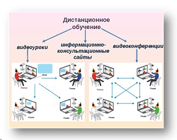
Влияние общества на человека
Приготовление дезинфицирующих растворов различной концентрации Практические работы по географии для 6 класса Организация работы процедурного кабинета Изменения в неживой природе осенью Уборка процедурного кабинета Сольфеджио. Все правила по сольфеджио Балочные системы. Определение реакций опор и моментов защемления |
Draw a diagram of the structure of plasmid pBR322Содержание книги
Поиск на нашем сайте
Draw a diagram of an experiment in genetic engineering (design recDNA) and give a description of the main stages Molecular cloning is the laboratory process used to create recombinant DNA. It is one of two widely used methods (along withpolymerase chain reaction, abbr. PCR) used to direct the replication of any specific DNA sequence chosen by the experimentalist. The fundamental difference between the two methods is that molecular cloning involves replication of the DNA within a living cell, while PCR replicates DNA in the test tube, free of living cells. Formation of recombinant DNA requires a cloning vector, a DNA molecule that will replicate within a living cell. Vectors are generally derived from plasmids or viruses, and represent relatively small segments of DNA that contain necessary genetic signals for replication, as well as additional elements for convenience in inserting foreign DNA, identifying cells that contain recombinant DNA, and, where appropriate, expressing the foreign DNA. The choice of vector for molecular cloning depends on the choice of host organism, the size of the DNA to be cloned, and whether and how the foreign DNA is to be expressed.[5] The DNA segments can be combined by using a variety of methods, such as restriction enzyme/ligase cloning or Gibson assembly. In standard cloning protocols, the cloning of any DNA fragment essentially involves seven steps: (1) Choice of host organism and cloning vector, (2) Preparation of vector DNA, (3) Preparation of DNA to be cloned, (4) Creation of recombinant DNA, (5) Introduction of recombinant DNA into the host organism, (6) Selection of organisms containing recombinant DNA, (7) Screening for clones with desired DNA inserts and biological properties.
Construction of recombinant DNA, in which a foreign DNA fragment is inserted into a plasmid vector. In this example, the gene indicated by the white color is inactivated upon insertion of the foreign DNA fragment.
Microinjection
Xenopus oocytes have been widely used for the study of transcription by microinjection because oocytes contain between 6,000 and 100,000 or more RNA polymerase molecules than somatic cells. Microinjection is technically easy because of large size of oocytes. Some of the endogenous pattern of gene regulation during development has been characterized (Wickens and Laskey, 1981). The injected DNA integrates randomly with nuclear DNA and its expression could be possible only when the foreign DNA is attached to a suitable promoter sequence. 73 Basic properties of cryoprotectors and their components A cryoprotectant is a substance that is used to protect biological tissue from freezing damage (i.e. that due to ice formation). Without protection, cells will rupture when they freeze as a result of expanding water, causing severe injury or death to living organisms, and ruining tissue samples or frozen food products. The compound can work in a number of different ways. A common approach is to lower the freezing point, keeping the tissue flexible at temperatures that would normally result in freezing. Others bond to specific molecules to help tissue retain its structure under the intense pressures of cold temperatures. Cryoprotectants must (1) easily penetrate cells, and (2) not be toxic to the cell. Conventional cryoprotectants are glycols (alcohols containing at least two hydroxyl groups), such as ethylene glycol[citation needed], propylene glycol, and glycerol. Ethylene glycol is commonly used as automobile antifreeze, and propylene glycol has been used to reduce ice formation in ice cream. Dimethyl sulfoxide (DMSO) is also regarded as a conventional cryoprotectant. Glycerol and DMSO have been used for decades by cryobiologists to reduce ice formation in sperm and embryos that are cold-preserved in liquid nitrogen. Mixtures of cryoprotectants have less toxicity and are more effective than single-agent cryoprotectants. A mixture of formamide with DMSO (dimethyl sulfoxide), propylene glycol, and a colloid was for many years, the most effective of all artificially created cryoprotectants. Cryoprotectant mixtures have been used for vitrification (i.e. solidification without crystal ice formation). Vitrification has important applications in preserving embryos, biological tissues, and organs for transplant. Vitrification is also used in cryonics in an effort to eliminate freezing damage. Some cryoprotectants function by lowering the glass transition temperature of a solution or of a material. In this way, the cryoprotectant prevents actual freezing, and the solution maintains some flexibility in a glassy phase. Many cryoprotectants also function by forming hydrogen bonds with biological molecules as water molecules are displaced. Hydrogen bonding in aqueous solutions is important for proper protein and DNA function. Thus, as the cryoprotectant replaces the water molecules, the biological material retains its native physiological structure and function, although they are no longer immersed in an aqueous environment. This preservation strategy is most often utilized in anhydrobiosis.
|
||||||||||
|
|
Последнее изменение этой страницы: 2016-08-14; просмотров: 382; Нарушение авторского права страницы; Мы поможем в написании вашей работы! infopedia.su Все материалы представленные на сайте исключительно с целью ознакомления читателями и не преследуют коммерческих целей или нарушение авторских прав. Обратная связь - 216.73.216.33 (0.006 с.) |