Заглавная страница Избранные статьи Случайная статья Познавательные статьи Новые добавления Обратная связь FAQ Написать работу КАТЕГОРИИ: ТОП 10 на сайте Приготовление дезинфицирующих растворов различной концентрацииТехника нижней прямой подачи мяча. Франко-прусская война (причины и последствия) Организация работы процедурного кабинета Смысловое и механическое запоминание, их место и роль в усвоении знаний Коммуникативные барьеры и пути их преодоления Обработка изделий медицинского назначения многократного применения Образцы текста публицистического стиля Четыре типа изменения баланса Задачи с ответами для Всероссийской олимпиады по праву 
Мы поможем в написании ваших работ! ЗНАЕТЕ ЛИ ВЫ?
Влияние общества на человека
Приготовление дезинфицирующих растворов различной концентрации Практические работы по географии для 6 класса Организация работы процедурного кабинета Изменения в неживой природе осенью Уборка процедурного кабинета Сольфеджио. Все правила по сольфеджио Балочные системы. Определение реакций опор и моментов защемления |
Objects and methods of animal biotechnologyСодержание книги
Поиск на нашем сайте
Objects and methods of animal biotechnology Animal biotechnology is the use of science and engineering to modify living organisms. The goal is to make products, to improve animals and to develop organisms for specific agricultural and medicine uses. Animal Biotechnology methods and tools are based on methods of: ü Developmental Biology-is the study of the process by which organisms grow and develop. Modern developmental biology studies the genetic control of cell growth, differentiation and morphogenesis, which is the process that gives rise to tissues, organs and anatomy, and even regeneration and aging,[1] more recently. ü Cell Biology - is a scientific discipline that studies cells – their physiological properties, their structure, the organelles they contain, interactions with their environment, their life cycle, division and death. This is done both on amicroscopic and molecular level. Cell biology research encompasses both the great diversity of single-celled organisms like bacteria andprotozoa, as well as the many specialized cells in multicellular organisms such as humans, plants, and sponges. ü Genetics and Molecular Biology - is the branch of biology that deals with the molecular basis of biological activity. This field overlaps with other areas of biology and chemistry, particularlygenetics and biochemistry. Molecular biology chiefly concerns itself with understanding the interactions between the various systems of a cell, including the interactions between the different types of DNA, RNA and protein biosynthesis as well as learning how these interactions are regulated. ü Gene Engineering - is the direct manipulation of an organism's genome using biotechnology. (Indirect genetic modification through artificial selection has been practiced for centuries.) New DNA may be inserted in the host genome by first isolating and copying the genetic material of interest using molecular cloning methods to generate a DNA sequence, or by synthesizing the DNA, and then inserting this construct into the host organism. Genes may be removed, or "knocked out", using a nuclease. Gene targeting is a different technique that uses homologous recombinationto change an endogenous gene, and can be used to delete a gene, remove exons, add a gene, or introduce point mutations. Objects: Laboratory animals ü Mouse ü Drosophila ü Chinese hamster ü Sea urchin ü Xenopus ü Rat ü Rabbit Farm animals ü cattle (cows, buffalo) ü small ruminants (sheep and goats) ü horses ü pigs ü poultry (chicken, turkey, duck, goose) ü fish (salmon, carp, herring) ü silkworm etc. Improving human health depends on the understanding of inside biology (through genetics, biochemistry, physiology, immunology, anatomy etc) as well as outside (through environmental contacts with other living and non-living things/products). For this purpose, many human diseases (e.g. genetic, acquired, metabolic or infectious) are modeled in animals to develop diagnostic assays, test therapies and preclinical research on scientific basis. With advances in biotechnology, more and more assays and preclinical trials are conducted in the animal model systems to understand the disease and functional biology because animal models mimic the human biology very closely. Popular lab animals include mice, rats, rabbits, fish and Drosophila. It is important to learn general biology and handling skills for these animals to use them inexperimental research.This is why the use of animals continue to be mandatory to meet the statutory requirements. -Drosophila, usually the species Drosophila melanogaster - a kind of fruit fly, famous as the subject of genetics experiments by Thomas Hunt Morgan and others. Easily raised in lab, rapid generations, mutations easily induced, many observable mutations. Recently, Drosophila has been used for neuropharmacological research. -Rat (Rattus norvegicus) - particularly useful as a toxicology model; also particularly useful as a neurological model and source of primary cell cultures, owing to the larger size of organs and suborganellar structures relative to the mouse. -African clawed frog (Xenopus laevis) - eggs and embryos from these frogs are used in developmental biology, cell biology,biotech, toxicology, and neuroscience -fish - has a nearly transparent body during early development, which provides unique visual access to the animal's internal anatomy. The fishs are used to study development, toxicology and toxicopathology,specific gene function and roles of signaling pathways. -Mouse - the classic model vertebrate. Many inbred strains exist, as well as lines selected for particular traits, often of medical interest, e.g. body size, obesity, muscularity, voluntary wheel-running behavior. -monkey - used for studies on infectious disease and cognition. -Guinea pig - used by bacteriologists as a host for bacterial infections, hence a byword for "laboratory animal" even though less commonly used today. -Chicken - used for developmental studies, as it is an amniote and excellent for micromanipulation (e.g. tissue grafting) and over-expression of gene products -Cat - used in neurophysiological research -Dog - an important respiratory and cardiovascular model, also contributed to the discovery of classical conditioning.
The factors influenced on microclonal propagation in plant cell culture. Factors that influence micro clonal propagation: The factors that important roles in the degree of success achieved in a given micropropagation or plant regeneration system include
1. The genotype of the donor, 2. The physiological conditions of the donor material, 3. The explants source, 4. The orientation and size of explants in culture, 5. The culture medium composition (s), 6. Interactions of endogenous hormones with exogenously supplied growth regulators, 7. The incubation conditions (including light quality and Intensity, Temperature, Relative humidity and Air quality) and 8. The timing of the subculture interval/changes in medium / incubation treatment.
Techniques Culture of embryos can either be performed in an artificial culture medium or in an autologous endometrial coculture (on top of a layer of cells from the woman's own uterine lining). With artificial culture medium, there can either be the same culture medium throughout the period, or a sequential system can be used, in which the embryo is sequentially placed in different media. For example, when culturing to the blastocyst stage, one medium may be used for culture to day 3, and a second medium is used for culture thereafter.[2] Single or sequential medium are equally effective for the culture of human embryos to the blastocyst stage.[3] Artificial embryo culture media basically contain glucose, pyruvate, and energy-providing components, but the addition of amino acids, nucleotides, vitamins, and cholesterol improve the performance of embryonic growth and development.[4] Methods to permit dynamic embryo culture with fluid flow and embryo movement are also available.[5] A new method in development uses the uterus as an incubator and the naturally occurring intrauterine fluids as culture medium by encapsulating the embryos in permeable intrauterine vessel.
Morphological and functional features of gametes - eggs and sperm Size and shape The egg cell (or ovum, or oocyte) is the largest human cell. She measures 0.15 to 0.2 mm and is just visible to the naked eye. She is also the roundest cell, she is almost perfectly round (Fig. 4). She therefore has the largest volume in relation to her surface. The cell consists of a large amount of cytoplasm (= cell fluid) in which the nucleus is dissolved (and therefore invisible) until just before conception. Sperm cells are the smallest human cells. They are no more than a nucleus with a small amount of cytoplasm, some mitochondria (the energy suppliers of the cell) and a long tail. They have hardly any content and are the straightest cells.
Mobility In contrast, the ovum is externally not active. After her release, she is passively moved by the fluid-flow in the oviduct (uterine tube), while the sperm cells are active, using their tails to swim against the stream of fluid in the oviduct. They are externally active and mobile. The ovum is internally mobile and externally passive, this is a polarity. The sperm shows the opposite: internally passive and externally mobile. Egg cell and sperm have a polarity and are opposite to each other, we see a double polarity. Sperm cells do not absorb or release substances. There is no interaction with the environment. They live about 3 to 5 days in the womb and can be preserved and frozen at temperatures below 60 °C. They are not easy to destroy. They are closed off from the environment and metabolically passive. The open and vulnerable state of the egg cell is polar to the closed and robust state of the sperm cells. The ovum is alone and the sperm are with millions. One sperm cell is nothing, one ovum determines everything. One is polar to millions. One comprises everything, it is all there is, whereas the millions of sperm cells are infinitive, have no importance on their own. Location The ovum develops in warm- and sperm in relative cold conditions. Development Egg cells are old cells that became mature. Primordial oocytes are in a process of dying. Sperm cells are newly formed and are young. The maturation process of ova is an expiring process, it stops. The formation of the sperm is a vital process, it never stops. 37 Cell markers. Types of selective markers. Selectable marker genes, also known as marker genes or selectable markers, are genesequences which are intentionally introduced into cells as developmental tools inrecombinant DNA technology. This technology is used to transfer genes for desired traits between organisms. Selectable marker genes are important for the production of novel or new organisms. They are used as a tool to identify those cells which have successfully incorporated genes for a desired trait during transformation. Since the percentage of cells that will incorporate genetic material during transformation is low, selectable markers are extremely important because they act as "genetic tags" to easily identify successfully modified cells. However, they have no actual function in the novel organism. The Science of Selectable Marker Genes. Selectable markers can be used in bacterial, plant, animal or other cells. The following is a summary of how selectable markers are incorporated into novel organisms:1.Gene Construct - includes the genetic material for the desired trait and may include selectable markers, genetic promoters (genes which may aid the expression of the trait) and vectors (the instrument- e.g. a virus or plasmid, which carries new DNA into a cell). 2. Agent - the lethal substance to which the selectable marker is tolerant/resistant (e.g., antibiotics, herbicides, etc.). 3.Transformed Cells - those cells that have incorporated the gene construct. Types of Selectable Marker Genes. There are four categories of selectable marker genes presently used as tools to develop novel products. The different types of selectable markers and their respective modes of action are described below. Antibiotic Resistant Marker Genes. Antibiotic resistant marker genes confer the trait of resistance to a specific antibiotic. For example, the neomycin phosphotransferase-II (NPT-II) gene, is a selectable marker gene that is the blueprint for resistance to the antibiotics neomycin and kanamycin. While they are useful in the early development of transgenic cells, antibiotic resistant marker genes are engineered to be expressed at minimal and often undetectable levels in the final product. Herbicide Tolerant Marker Genes. Herbicide tolerant marker genes confer the trait of tolerance to the application of a specific herbicide. For instance, the gene conferring tolerance to the herbicide glufosinate ammonium is often used as a selectable marker in plant biotechnology. Those cells which survive the exposure to the herbicide are selected and regenerated into whole organisms. Metabolic/Auxotrophic Marker Genes. Metabolic or auxotrophic marker genes enable transformed cells to synthesize an essential component, usually an amino acid, which the cells cannot otherwise produce. The surrounding medium is made to intentionally lack the essential component, which cells require to grow. Cells that have successfully incorporated the selectable marker and the rest of the gene construct will produce the essential components within the cells, and thereby survive. These cells are selected and regenerated into whole organisms. Screenable Marker Genes. Screenable markers, also known as assayable markers, are genes which encode for a protein that can be identified through various laboratory assessments. The presence of the protein confirms that transformation has taken place. Screenable markers are not commonly used because their use is very time consuming, expensive equipment and supplies are required to perform the assessments and the final tissue product has to be destroyed to determine if transformation has taken place. Some of these selectable marker genes are more commonly used than others because they are better understood and are more compatible with the tissues/cells under transformation. For example, herbicide tolerance is a selectable marker commonly used in the development of plants with novel traits because the mode of action of herbicide tolerance has been extensively studied and it is compatible with plant tissue and cells. Removing Selectable Marker Genes. Selective breeding and cellular and molecular biology techniques are used to remove the selectable marker genes from novel cells and organisms while retaining the genes of commercial interest. Of these techniques, the selective breeding of novel organisms to breed out the selectable marker is the most common. 39) Basic approaches and principles of gene therapy. Gene therapy ex vivo, in vivo, in situ. Gene therapy is the use of DNA as a pharmaceutical agent to treat disease. It derives its name from the idea that DNA can be used to supplement or alter genes within an individual's cells as a therapy to treat disease. The most common form of gene therapy involves using DNA that encodes a functional, therapeutic gene to replace a mutated gene. Other forms involve directly correcting a mutation, or using DNA that encodes a therapeutic protein drug (rather than a natural human gene) to provide treatment. In gene therapy, DNA that encodes a therapeutic protein is packaged within a "vector", which is used to get the DNA inside cells within the body. Once inside, the DNA becomes expressed by the cell machinery, resulting in the production of therapeutic protein, which in turn treats the patient's disease. Gene therapy may be classified into the two following types: [edit] Somatic gene therapy In somatic gene therapy, the therapeutic genes are transferred into the somatic cells, or body, of a patient. Any modifications and effects will be restricted to the individual patient only, and will not be inherited by the patient's offspring or later generations. Somatic gene therapy represents the mainstream line of current basic and clinical research, where the therapeutic DNA transgene (either integrated in the genome or as an external episome or plasmid) is used to treat a disease in an individual. [edit] Germ line gene therapy In germ line gene therapy, germ cells, i.e., sperm or eggs, are modified by the introduction of functional genes, which are integrated into their genomes. This would allow the therapy to be heritable and passed on to later generations. Although this should, in theory, be highly effective in counteracting genetic disorders and hereditary diseases, many jurisdictions prohibit this for application in human beings, at least for the present, for a variety of technical and ethical reasons This can be accomplished in three ways: ex vivo, in situ, or in vivo. Ex vivo involves removing cells from the patient, altering the genetic material, and placing them back into the patient. In situ requires the vector be placed directly into the affected tissues. In vivo gene therapy involves injecting the vector into the bloodstream.
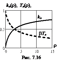
Draw a diagram of an experiment in genetic engineering (design recDNA) and give a description of the main stages Molecular cloning is the laboratory process used to create recombinant DNA. It is one of two widely used methods (along withpolymerase chain reaction, abbr. PCR) used to direct the replication of any specific DNA sequence chosen by the experimentalist. The fundamental difference between the two methods is that molecular cloning involves replication of the DNA within a living cell, while PCR replicates DNA in the test tube, free of living cells. Formation of recombinant DNA requires a cloning vector, a DNA molecule that will replicate within a living cell. Vectors are generally derived from plasmids or viruses, and represent relatively small segments of DNA that contain necessary genetic signals for replication, as well as additional elements for convenience in inserting foreign DNA, identifying cells that contain recombinant DNA, and, where appropriate, expressing the foreign DNA. The choice of vector for molecular cloning depends on the choice of host organism, the size of the DNA to be cloned, and whether and how the foreign DNA is to be expressed.[5] The DNA segments can be combined by using a variety of methods, such as restriction enzyme/ligase cloning or Gibson assembly. In standard cloning protocols, the cloning of any DNA fragment essentially involves seven steps: (1) Choice of host organism and cloning vector, (2) Preparation of vector DNA, (3) Preparation of DNA to be cloned, (4) Creation of recombinant DNA, (5) Introduction of recombinant DNA into the host organism, (6) Selection of organisms containing recombinant DNA, (7) Screening for clones with desired DNA inserts and biological properties.
Construction of recombinant DNA, in which a foreign DNA fragment is inserted into a plasmid vector. In this example, the gene indicated by the white color is inactivated upon insertion of the foreign DNA fragment.
Microinjection
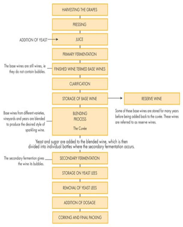
Xenopus oocytes have been widely used for the study of transcription by microinjection because oocytes contain between 6,000 and 100,000 or more RNA polymerase molecules than somatic cells. Microinjection is technically easy because of large size of oocytes. Some of the endogenous pattern of gene regulation during development has been characterized (Wickens and Laskey, 1981). The injected DNA integrates randomly with nuclear DNA and its expression could be possible only when the foreign DNA is attached to a suitable promoter sequence. 73 Basic properties of cryoprotectors and their components A cryoprotectant is a substance that is used to protect biological tissue from freezing damage (i.e. that due to ice formation). Without protection, cells will rupture when they freeze as a result of expanding water, causing severe injury or death to living organisms, and ruining tissue samples or frozen food products. The compound can work in a number of different ways. A common approach is to lower the freezing point, keeping the tissue flexible at temperatures that would normally result in freezing. Others bond to specific molecules to help tissue retain its structure under the intense pressures of cold temperatures. Cryoprotectants must (1) easily penetrate cells, and (2) not be toxic to the cell. Conventional cryoprotectants are glycols (alcohols containing at least two hydroxyl groups), such as ethylene glycol[citation needed], propylene glycol, and glycerol. Ethylene glycol is commonly used as automobile antifreeze, and propylene glycol has been used to reduce ice formation in ice cream. Dimethyl sulfoxide (DMSO) is also regarded as a conventional cryoprotectant. Glycerol and DMSO have been used for decades by cryobiologists to reduce ice formation in sperm and embryos that are cold-preserved in liquid nitrogen. Mixtures of cryoprotectants have less toxicity and are more effective than single-agent cryoprotectants. A mixture of formamide with DMSO (dimethyl sulfoxide), propylene glycol, and a colloid was for many years, the most effective of all artificially created cryoprotectants. Cryoprotectant mixtures have been used for vitrification (i.e. solidification without crystal ice formation). Vitrification has important applications in preserving embryos, biological tissues, and organs for transplant. Vitrification is also used in cryonics in an effort to eliminate freezing damage. Some cryoprotectants function by lowering the glass transition temperature of a solution or of a material. In this way, the cryoprotectant prevents actual freezing, and the solution maintains some flexibility in a glassy phase. Many cryoprotectants also function by forming hydrogen bonds with biological molecules as water molecules are displaced. Hydrogen bonding in aqueous solutions is important for proper protein and DNA function. Thus, as the cryoprotectant replaces the water molecules, the biological material retains its native physiological structure and function, although they are no longer immersed in an aqueous environment. This preservation strategy is most often utilized in anhydrobiosis. Cryopreservation of gametes Spermatozoa Typically the process of preserving male gametes is more straightforward than that of preserving female gametes, partly because a single semen sample typically contains millions of sperm, so even modest cryosurvival rates result in large quantities of sperm. There are however a number of factors which influence the effectiveness of sperm cryopreservation including: Quality of the semen sample - The proportion of live sperm which survive the freezing and thawing process is higher in a normozoospermic sample (24.9% survival) than in a oligoozoospermic sample (11.9% survival). Semen preparation - The method by which semen is washed prior to freezing also influences post-thaw vitality. In normozoospermic semen samples, the proportion of sperm which survive cryopreservation increases from 24.9% with no preparation, to 35.6% if sperm are washed using a ‘swim up' technique prior to cryopreservation. Both the swim up and density gradient centrifugation techniques also significantly improve the survival rate of spermatozoa in oligozoospermic samples (from 11.9% when not washed to 27.7% using swim up and 22.4% using centrifugation). Oocytes The cryopreservation of oocytes has proven more challenging than the cryopresevation of sperm, in particular because many regimens cause damage to an egg's zona pellucida (i.e. shell). However there are now a range of cryopreservation protocols which can be used to preserve mature or immature oocytes. While survival rates for immature oocytes have reached 60%, the freezing process often alters the oocyte development process and fertilisation and live birth rates for surviving oocytes, are consistently lower than those achieved using fresh oocytes. Cryopreservation of embryos Overall cryopreservation of embryos results in about 5% chance of live birth, while for blastocysts the rate is around 10%. It is considerably more efficient than the cryopreservation of oocytes and it is therefore most common for oocytes to be fertilised in vitro prior to cryopreservation. Reproductive outcomes following embryo cryopreservation depend on the stage of embryo development, both at the time of freezing and at the time of implantation. Embryos which have recommenced mitosis (division and replication which leads to the growth of the embryo) post-thaw but pre-implantation have a higher chance of implantation. Cryopreservation of ovarian and testicular tissues The cryopreservation of gonadal tissues is most commonly performed when a patient must immediately undergo toxic treatment which threatens their fertility. As the urgency of such treatment may not allow time for the collection of mature gametes (e.g. because there is no time to perform ovarian stimulation or because they have not yet gone through puberty), removal and storage of gonadal tissues which contain immature gametes is sometimes the only option for fertility preservation. Wine production
85 The microorganisms used in industry The use of microorganism in large scale production of food and industrial products is being done worldwide.The sources of food production in such cases may be animals or plants but the processing is done by enzymatic activities by microorganism only. Microorganism contains various enzymes which are capable of degradation of substrates. This is also known as fermentation process in which the degradations is not completed and results in useful by products. These by products include, beverages, antibiotics, milk by products etc which are used by humans as nutritive foods. With the development of technology like genetic engineering, many mutants are developed which are capable of performing extra with respect to production quality and quantity as compared to their wild types. Today pharmaceutical agents like antibiotics and other drugs are manufactured at large scale which utilizes microorganism. The history of microbiology has given us very broad spectrum antibiotics like Penicillin and Streptomycin which are still in use at large throughout the globe. Foods which are originated from animals are enzymatic ally processed by specific microorganisms resulting in increase in their nutritive value. These foods are fermented foods like Yogurt, milk by products like cheese, sweet chocolates and silage. Many algae are today used as source of protein. Fungus like mushroom is today being used as source of nutrition as well as medicine. Bacteria like Lactic acid bacteria are used in production of pure curd and other milk products. Bacteria like Bifidobacteria are being used in food industry as probiotics which helps in curing of diseases of digestive systems and intestinal disorders. Polysaccharides, polyamides, polyesters and many other varieties of biopolymers are produced by many microorganisms. These are ranging from plastics to viscous solutions. Today many researches in drug delivery and tissue engineering are being successfully done with the help of genetically manipulated microorganisms which are producing biopolymers which are having medical applications. Many wild as well as genetically mutated strains are used in industries for biosynthesis of cellulose, levan, hyaluronic acids, polysaccharides and organic acids, etc. Today many pollutants are degraded with the help of saprophytes (specific type of bacteria/fungi capable of surviving in waste organic material their by metabolism) this process is also known as biodegradation. Today bioremediation and other methods like biotransformation are used for cleaning of the environments. Today even heavy metals like mercury which is toxic and results in biomagnifications. The degradation of this is very costly by chemical and other standards technologies. Therefore the alternative method is bioremediation. Today in modern societies, lot of waste is being generated from domestic wastes. These are accumulating every day and are very harmful to not only the society but also the environment (Our mother earth). The processing of such waste using living organisms is known as biotreatment. These methods are helping society and saving earth from accumulation of hazardous wastes. This method of using microorganisms in degradation of hazardous waste is not only useful but also simple, cost effective and eco friendly. The systematic method of biotreatment is done with the help of bioreactors having aeration system, baffles which suitable for microbial enzymatic reaction. Apart from waste treatments, microorganisms are used in production of biological like insulin, serum antibodies, and essential hormones. Today with the development of technology and science, new ways of diagnosis of diseases are being used for early detection using microorganism’s e.g. rapid microbial test, enzyme employed detection, etc. One of the microbe known as Clostridium is useful in treatment of malignant cells like cancers. These organisms have the ability to selectively target cancerous cells. Streptomyces genus is used worldwide in the production of medicines and agriculture. These microorganisms process many secondary metabolites like antibiotics and plant growth hormones like Gibberilla (Microorganism used is Gibberella fujikuroi).
Commercial Applications Yeast has long been considered to be the organism of choice for the production of alcoholic beverages, bread, and a large variety of industrial products. This is based on the ease with which the metabolism of yeast can be manipulated using genetic techniques, the speed with which it can be grown to high cell yields (biomass), the ease with which this biomass can be separated from products and the knowledge that it is generally recognized as safe (GRAS).
Objects and methods of animal biotechnology Animal biotechnology is the use of science and engineering to modify living organisms. The goal is to make products, to improve animals and to develop organisms for specific agricultural and medicine uses. Animal Biotechnology methods and tools are based on methods of: ü Developmental Biology-is the study of the process by which organisms grow and develop. Modern developmental biology studies the genetic control of cell growth, differentiation and morphogenesis, which is the process that gives rise to tissues, organs and anatomy, and even regeneration and aging,[1] more recently. ü Cell Biology - is a scientific discipline that studies cells – their physiological properties, their structure, the organelles they contain, interactions with their environment, their life cycle, division and death. This is done both on amicroscopic and molecular level. Cell biology research encompasses both the great diversity of single-celled organisms like bacteria andprotozoa, as well as the many specialized cells in multicellular organisms such as humans, plants, and sponges. ü Genetics and Molecular Biology - is the branch of biology that deals with the molecular basis of biological activity. This field overlaps with other areas of biology and chemistry, particularlygenetics and biochemistry. Molecular biology chiefly concerns itself with understanding the interactions between the various systems of a cell, including the interactions between the different types of DNA, RNA and protein biosynthesis as well as learning how these interactions are regulated. ü Gene Engineering - is the direct manipulation of an organism's genome using biotechnology. (Indirect genetic modification through artificial selection has been practiced for centuries.) New DNA may be inserted in the host genome by first isolating and copying the genetic material of interest using molecular cloning methods to generate a DNA sequence, or by synthesizing the DNA, and then inserting this construct into the host organism. Genes may be removed, or "knocked out", using a nuclease. Gene targeting is a different technique that uses homologous recombinationto change an endogenous gene, and can be used to delete a gene, remove exons, add a gene, or introduce point mutations. Objects: Laboratory animals ü Mouse ü Drosophila ü Chinese hamster ü Sea urchin ü Xenopus ü Rat ü Rabbit Farm animals ü cattle (cows, buffalo) ü small ruminants (sheep and goats) ü horses ü pigs ü poultry (chicken, turkey, duck, goose) ü fish (salmon, carp, herring) ü silkworm etc. Improving human health depends on the understanding of inside biology (through genetics, biochemistry, physiology, immunology, anatomy etc) as well as outside (through environmental contacts with other living and non-living things/products). For this purpose, many human diseases (e.g. genetic, acquired, metabolic or infectious) are modeled in animals to develop diagnostic assays, test therapies and preclinical research on scientific basis. With advances in biotechnology, more and more assays and preclinical trials are conducted in the animal model systems to understand the disease and functional biology because animal models mimic the human biology very closely. Popular lab animals include mice, rats, rabbits, fish and Drosophila. It is important to learn general biology and handling skills for these animals to use them inexperimental research.This is why the use of animals continue to be mandatory to meet the statutory requirements. -Drosophila, usually the species Drosophila melanogaster - a kind of fruit fly, famous as the subject of genetics experiments by Thomas Hunt Morgan and others. Easily raised in lab, rapid generations, mutations easily induced, many observable mutations. Recently, Drosophila has been used for neuropharmacological research. -Rat (Rattus norvegicus) - particularly useful as a toxicology model; also particularly useful as a neurological model and source of primary cell cultures, owing to the larger size of organs and suborganellar structures relative to the mouse. -African clawed frog (Xenopus laevis) - eggs and embryos from these frogs are used in developmental biology, cell biology,biotech, toxicology, and neuroscience -fish - has a nearly transparent body during early development, which provides unique visual access to the animal's internal anatomy. The fishs are used to study development, toxicology and toxicopathology,specific gene function and roles of signaling pathways. -Mouse - the classic model vertebrate. Many inbred strains exist, as well as lines selected for particular traits, often of medical interest, e.g. body size, obesity, muscularity, voluntary wheel-running behavior. -monkey - used for studies on infectious disease and cognition. -Guinea pig - used by bacteriologists as a host for bacterial infections, hence a byword for "laboratory animal" even though less commonly used today. -Chicken - used for developmental studies, as it is an amniote and excellent for micromanipulation (e.g. tissue grafting) and over-expression of gene products -Cat - used in neurophysiological research -Dog - an important respiratory and cardiovascular model, also contributed to the discovery of classical conditioning.
|
||||||||||||
|
|
Последнее изменение этой страницы: 2016-08-14; просмотров: 385; Нарушение авторского права страницы; Мы поможем в написании вашей работы! infopedia.su Все материалы представленные на сайте исключительно с целью ознакомления читателями и не преследуют коммерческих целей или нарушение авторских прав. Обратная связь - 216.73.216.20 (0.01 с.) |