Заглавная страница Избранные статьи Случайная статья Познавательные статьи Новые добавления Обратная связь FAQ Написать работу КАТЕГОРИИ: ТОП 10 на сайте Приготовление дезинфицирующих растворов различной концентрацииТехника нижней прямой подачи мяча. Франко-прусская война (причины и последствия) Организация работы процедурного кабинета Смысловое и механическое запоминание, их место и роль в усвоении знаний Коммуникативные барьеры и пути их преодоления Обработка изделий медицинского назначения многократного применения Образцы текста публицистического стиля Четыре типа изменения баланса Задачи с ответами для Всероссийской олимпиады по праву 
Мы поможем в написании ваших работ! ЗНАЕТЕ ЛИ ВЫ?
Влияние общества на человека
Приготовление дезинфицирующих растворов различной концентрации Практические работы по географии для 6 класса Организация работы процедурного кабинета Изменения в неживой природе осенью Уборка процедурного кабинета Сольфеджио. Все правила по сольфеджио Балочные системы. Определение реакций опор и моментов защемления |
Ex. 1. Analyze the ing – forms and ed-forms and translate the sentences.Содержание книги
Поиск на нашем сайте
Chapter 8
1. Class work. Ex. 1. Analyze the ing – forms and ed-forms and translate the sentences.
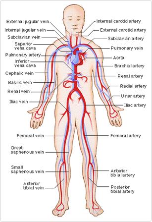
Ex.2. Memorize the new words. · the circulatory system = the cardiovascular system - сердечно – сосудистая система · to circulate – циркулировать · to circle – окружить · a circle - круг · to serve – обслуживать, служить · independently - независимо · in order to – для того, чтобы · to provide with - снабжать, to provide for - предусматривать · to supply – обеспечивать, снабжать · to convey = to deliver - транспортировать, передавать · an arch - дуга Ex. 3. Using your knowledge of anatomy translate the medical terms in picture 1. Ex. 4. Read, translate and memorize the basic blood vessel terminology: Basic Blood Vessel Terminology
Picture 1. The body's circulatory system.
Ex. 5. a) Look through text A. b) Find the ing – forms and ed- forms and analyze them. c) Read and translate the sentences with the ing – forms and ed-forms. d) Read the text again, divide it into logical units and entitle each unit. e) Make the plan of the text. Text A. The cardiovascular system The body's circulatory system really has three parts: pulmonary circulation, coronary circulation, and systemic circulation or the lungs (pulmonary), the heart (coronary), and the rest of the system (systemic). Each part is working independently in order to all work together. The heart, the lungs, and the blood vessels work together and form the circulatory system. The healthy function of the entire body's circulatory system is influenced by the good work of any organ listed above. The cardiovascular system consists of the heart and blood vessels. Together, these provide a continuous flow of blood to the body, supplying the tissues with oxygen and nutrients. The arteries carry blood away from the heart. Oxygenated blood is pumped out of the heart through the body's main artery — the aorta. The blood vessels carrying the oxygen-rich blood to the organs are known as arteries. Arteries transport blood throughout the body, supplying tissues with oxygen and nutrients which are needed by any living being. The veins carry blood back toward the heart. The blood vessels delivering blood towards the heart are known as veins. Tiny vessels called capillaries deliver deoxygenated blood into small veins called venules, which join when forming veins. Blood flows through the veins to the body's two main veins (the vena cavae), delivering the blood back into the heart. On average, the body has about 5 liters of blood traveling through it by way of the circulatory system, the pumping of the heart forcing the blood on its journey. Ex. 6. a) Read text B. b) Entitle it. c) Find the information which is not included in text A.
Text B. The heart and circulatory system make up the cardiovascular system. The heart works as a pump that pushes blood to the organs, tissues, and cells of the body. Blood delivers oxygen and nutrients to every cell and removes the carbon dioxide and waste products made by those cells. Blood is carried from the heart to the rest of the body through a complex network of arteries, arterioles, and capillaries. Blood is returned to the heart through venules and veins. If all the vessels of this network in the body were laid end-to-end, they would extend for about 60,000 miles (more than 96,500 kilometers), which is far enough to circle the earth more than twice! Home work. Ex. 7. Read, translate and memorize the word combinations: Pulmonary circulation, coronary circulation, systemic circulation, blood vessels, to work independently in order for them to all work together, to provide a continuous flow of blood to your body, to supply the tissues with oxygen and nutrients, to pump oxygenated blood to the heart through the body's main artery, to carry the oxygen-rich blood to the organs known as arteries, to convey blood towards the heart, to deliver deoxygenated blood into small veins called venules, to flow through the veins to the body's two main veins, to travel through the body by way of the circulatory system, to force the blood on its journey, to circle the body.
Class work Picture 2. Heart Anatomy.
The Heart Muscle.
Ex. 15. Memorize the words: · Hollow - полый · a fist - кулак · communication – сообщение, связь · a hole – отверстие, дыра · to bring in – вводить, вносить · to drain – дренировать, осушать, забирать влагу · to ensure – обеспечивать, гарантировать · to advance a disease – запустить болезнь · to advance an idea – выдвинуть идею · OTC = over the counter medicines – безрецептурные лекарства · prescription medicines – рецептурные лекарства · a pacemaker – вводитель ритма · to extract - выделять · to remove - удалять · to add – добавлять, in addition to - кроме · to speed up – ускорять, работать быстрее · to let - позволять Atria and Ventricles Weight, size and location Right Heart and Left Heart Text C. Heart Anatomy a. The heart is an inner hollow organ. The heart weighs between 7 and 15 ounces (200 to 425 grams) and is a little larger than the size of your fist. The heart is located between the lungs in the middle of the chest, behind and to the left of the breastbone (sternum). The base of the heart is against the third rib. Its apex is against the interspaces between the fifth and the sixth costal cartilages. b. The heart is divided into 4 heart chambers, the right and left ventricles and atria. The heart, made up mainly of cardiac muscle tissue, is an organ pumping blood to the whole of the body. The inside of the heart is divided into 4 chambers, 2 atria and 2 ventricles. c. The atria are the chambers that receive blood from circulation. The ventricles are thicker walled chambers, located below the atria and connected with them by openings covered by heart valves. Their function is to pump blood from the heart into circulation. d. The heart is divided into right and left sides by a septum. Each side consists of one atrium and one ventricle. So the chambers of the heart are named:
The right side of the heart is responsible for pulmonary circulation to the lungs, the left side of the heart dealing with systemic circulation or circulation to the whole body. Normally, there is no communication between the right and left sides of the heart. Communications are present in abnormal conditions like atrial and ventricular septal defects, conditions that commonly go by the name ‘a hole in the heart’. e. The right atrium receives venous blood from the whole of the body. It has the superior vena cava and inferior vena cava opening into its cavity. While the superior vena cava brings in venous blood from the upper limbs, head and neck regions and walls of the chest and upper abdomen, the inferior vena cava drains the abdomen and the lower limbs. The blood entering the right atrium is deoxygenated – that is, its oxygen has been used up by the tissues of the body and this blood is rich in carbon dioxide. The right ventricle is almost triangular in shape. It opens into the pulmonary artery. The deoxygenated blood in the right ventricle is carried by the pulmonary artery to the lungs for the process of gas exchange, where carbon dioxide is exchanged for oxygen. f. The left atrium contains the openings of the superior and inferior pulmonary veins above. These blood vessels bring oxygenated blood from the lungs into the left atrium. When the left atrium contracts, the oxygen-rich blood in it is pumped into the left ventricle. The left ventricle is larger and more muscular than the right ventricle. The left ventricle receives oxygen-rich blood from the left atrium which it pumps into the aorta for circulation to the entire body. Heart and Palms. Recently, a medical science called dermatoglyphics has found that creases of the hands can help in the diagnosis of disease. The doctors discovered that baby’s palm print could often show serious heart defects. They believe that any genetic or external factors that damage the organs of the developing fetus can affect the fingers, palm or sole prints. And indeed, researchers have detected a number of congenital and hereditary disorders that leave their marks on the hand or feet. They collected the finger, palm and sole prints from 225 children diagnosed as having congenital heart defects and compared them with the prints of 400 persons without cardiac malformations. Finger and foot prints from both groups were the same. But the palm prints of the heart prints showed difference. In a normal person there is a point at which three creases near the wrist form a kind of three pointed star. But in nearly all the heart patients, however, it was not situated near the wrist, but higher in the palm, towards the fingers. Doctors note that in many cases children with heart defects may show no symptoms until years after birth – and then it is often too late for successful surgery. The researchers consider that hand prints should be used as a routine screening test for congenital cardiac disease in all babies. Home work. Ex. 19. Read, translate and memorize the word combinations: To be a little larger than the size of your fist, to be divided into 4 heart chambers, for effective pumping of blood, to pump blood from the heart into circulation, to be made up mainly of cardiac muscle tissue, to receive blood from circulation, to be almost triangular in shape, to be carried by the pulmonary artery to the lungs for the process of gas exchange, to contain the openings of the superior and inferior pulmonary veins above. Ex. 20. Match the medical terms with the sentences:
Class work. Text E. The heart is located between the lungs in the middle of the chest, behind and to the left of the breastbone (sternum). The base of the heart is against the third rib, its apex being against the interspace between the fifth and the sixth costal cartilages. A double-layered membrane called the pericardium surrounds the heart like a sac. The outer layer of the pericardium surrounds the roots of the heart’s major blood vessels and is attached by ligaments to the spinal column, diaphragm, and other parts of the body. The inner layer of the pericardium is attached to the heart muscle. A coating of fluid separates the two layers of membrane, letting the heart move as it beats, yet still be attached to your body. The heart has 4 chambers. The upper chambers are called the left and right atria, and the lower chambers are called the left and right ventricles. A wall of muscle called the septum separates the left and right atria and the left and right ventricles. The left ventricle is the largest and strongest chamber in the heart. The left ventricle’s chamber walls are only about a half-inch thick, but they have enough force to push blood through the aortic valve and into your body. The healthy function of the heart is influenced by the good work of any chamber. By the end of a long life, a person’s heart beats about (expanded and contracted) more than 3.5 billion times. In fact, each day, the average heart beats 100,000 times, pumping about 2,000 gallons (7,571 liters) of blood. Ex. 28. a) Translate the title of the article. b) Read the article and retell it in Russian.
A Tale of Creased Earlobes. Doctors know that anyone with high blood pressure, an elevated level of cholesterol in the blood and a big smoking habit runs a greater than average risk of a heart disease. Now heart specialists have found another very important prognostic sign – creased earlobes. The crease runs from the lowest corner of the ear-opening diagonally down the lobe. Its potential significance was first observed in 1960s by doctors working in Air Force hospital. They noticed that a high proportion of young airmen admitted to the coronary care unit had creased lobes and later concluded that they might be a sign of a heart disease. Only one of the eleven who did not have the crease had a heart trouble. The crease may be a good clue for the doctors. But in the absence of such established symptoms as chronic chest pains, a person who finds that his lobes are creased shouldn’t worry. Home work.
Text F. All of the body’s organs require blood for a good function. Blood is composed of a liquid portion known as plasma and several cell components. The cell components are red blood cells, white blood cells and platelets. The red blood cells are the cells that carry oxygen to the body’s organs. The blood vessels that carry the oxygen-rich blood to the organs are known as arteries. During the process of metabolism the organs develop carbon dioxide. As the oxygen-rich blood passes through the organ, the oxygen in the red blood cells is exchanged for the carbon dioxide. Blood rich in oxygen is bright red, whereas (в то время как) blood that has had the oxygen extracted is of a dark red to purple (пурпурный) color. The blood that has had its oxygen extracted is known as de-saturated (saturated – насыщенный) blood. The blood vessels that carry the de-saturated blood away from the organ are known as veins. The heart pumps nonstop and supplies your body with blood that is rich in oxygen. For its job, your heart needs its own oxygen-rich blood supply. Blood gets oxygen from the lungs and travels to your heart through the coronary arteries. A large vessel called the aorta carries blood from your heart to the rest (остальной) of your body. The coronary arteries branch off from the aorta. Smaller arteries branch off from the coronary arteries. These smaller arteries run over and through the heart muscle. They nourish (питать) every part of the heart with blood. Picture 4 Ex. 35. a) Read text G and entitle it. b) Answer the questions below. Find your answers in the text.
Home work. Class work. Home work. Chapter 8
1. Class work. Ex. 1. Analyze the ing – forms and ed-forms and translate the sentences.
|
||||||||||||||||||||||
|
|
Последнее изменение этой страницы: 2016-08-01; просмотров: 321; Нарушение авторского права страницы; Мы поможем в написании вашей работы! infopedia.su Все материалы представленные на сайте исключительно с целью ознакомления читателями и не преследуют коммерческих целей или нарушение авторских прав. Обратная связь - 216.73.216.33 (0.008 с.) |

 Ex. 14. Read, translate and memorize the basic heart terminology. It will help you to understand the basic terms related to the heart.