Заглавная страница Избранные статьи Случайная статья Познавательные статьи Новые добавления Обратная связь FAQ Написать работу КАТЕГОРИИ: ТОП 10 на сайте Приготовление дезинфицирующих растворов различной концентрацииТехника нижней прямой подачи мяча. Франко-прусская война (причины и последствия) Организация работы процедурного кабинета Смысловое и механическое запоминание, их место и роль в усвоении знаний Коммуникативные барьеры и пути их преодоления Обработка изделий медицинского назначения многократного применения Образцы текста публицистического стиля Четыре типа изменения баланса Задачи с ответами для Всероссийской олимпиады по праву 
Мы поможем в написании ваших работ! ЗНАЕТЕ ЛИ ВЫ?
Влияние общества на человека
Приготовление дезинфицирующих растворов различной концентрации Практические работы по географии для 6 класса Организация работы процедурного кабинета Изменения в неживой природе осенью Уборка процедурного кабинета Сольфеджио. Все правила по сольфеджио Балочные системы. Определение реакций опор и моментов защемления |
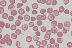
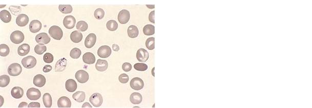
Image S5: ovalocytes and elliptocytesСодержание книги
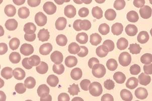
Поиск на нашем сайте Image S1: acanthocytes
Abetalipoproteinaemia - typical hyperchromic cells with projections of variable length, thickness and shape
J. Burthem, M. Brereton
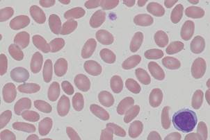
Three bite cells
J. Burthem, M. Brereton
G6PD deficiency (drug induced haemolysis) - frequent blister cells with associated damaged cells
J. Burthem, M. Brereton
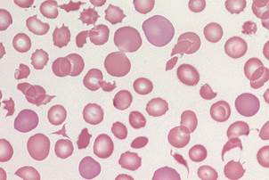
Image S4: echinocytes
Renal failure – cells with evenly spaced, short, blunt projections
J. Burthem, M. Brereton 2
Supplementary images: ICSH Recommendations for Peripheral Blood Cell Morphology
Standardization and Grading
Image S5: ovalocytes and elliptocytes
Hereditary elliptocytosis
J. Burthem, M. Brereton
Image S6: irregularly contracted cells
Unstable haemoglobin
J. Burthem, M. Brereton
Image S7: schistocytes
Thrombotic thrombocytopenic purpura – a wide range of fragmented red cells with polychromatic cells and other damaged cells. Platelets are absent from the film
J. Burthem, M. Brereton
Image S8: sickle cells
Sickle cell disease – typical forms together with other features, notably polychromasia, target cells, and a spherocytic cell.
J. Burthem, M. Brereton 3
Supplementary images: ICSH Recommendations for Peripheral Blood Cell Morphology
Standardization and Grading
Image S9: spherocytes
Autoimmune haemolytic anaemia – typical round, dense red cells
J. Burthem, M. Brereton
Image S10: stomatocytes
Hereditary stomatocytosis
J. Burthem, M. Brereton
Image S11: target cells
Haemoglobin C disease – target cells in association with irregularly contracted cells
J. Burthem, M. Brereton
Image S12: tear drop cells
Myelofibrosis
J. Burthem, M. Brereton 4
Supplementary images: ICSH Recommendations for Peripheral Blood Cell Morphology
Standardization and Grading
Image S13: basophilic stippling
Myelofibrosis - two stippled cells, one in teardrop form
J. Burthem, M. Brereton
Image S14: Howell-Jolly bodies
Auto-splenectomised patient with sickle cell disease
J. Burthem, M. Brereton
Image S15: Pappenheimer bodies
Sideroblastic anaemia – red cells contain small basophilic inclusions of variable size and shape in a limited cytoplasmic area
G. Rozenberg
(Copyright: Microscopic haematology: a practical guide for the laboratory 3e (c) 2011, Sydney, Elsevier Australia)
Image S16: nucleated red blood cell
Myelofibrosis – a typical late stage nucleated red cell in circulation
J. Burthem, M. Brereton 5
Supplementary images: ICSH Recommendations for Peripheral Blood Cell Morphology
Standardization and Grading
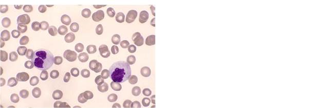
Image S18: Auer rods
AML – two blast cells containing relatively blunt-ended, single and multiple Auer rods
J. Burthem, M. Brereton
Image S19: hypergranulation
(neutrophils)
Hypergranular neutrophils post G-CSF treatment
J. Burthem, M. Brereton
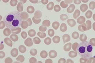
Image S20: hypogranulation
(neutrophils)
Myelodysplasia – hypogranular neutrophils.
Note also the atypical nuclear forms
G. Rozenberg
(Copyright: Microscopic haematology: a practical guide for the laboratory 3e (c) 2011, Sydney, Elsevier Australia) 6
Supplementary images: ICSH Recommendations for Peripheral Blood Cell Morphology
Standardization and Grading
Image S25: monoblasts
Acute monoblastic leukaemia – monoblasts and promonocytes
J. Burthem, M. Brereton
Image S28: hairy cells
Hairy cell leukaemia
J. Burthem, M. Brereton 8
Supplementary images: ICSH Recommendations for Peripheral Blood Cell Morphology
Standardization and Grading
Image S30: plasma cells
Plasma cell leukaemia. Note also the background protein staining and the associated red cell rouleaux. Note that one plasma cell has features of immaturity and may be regarded as a plasmablast.
J. Burthem, M. Brereton
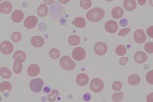
Image S33: giant platelets
Myelofibrosis – some large and giant abnormally granulated platelets and a micromegakaryocyte
J. Burthem, M. Brereton
Image S1: acanthocytes
Abetalipoproteinaemia - typical hyperchromic cells with projections of variable length, thickness and shape
J. Burthem, M. Brereton
Three bite cells
J. Burthem, M. Brereton
G6PD deficiency (drug induced haemolysis) - frequent blister cells with associated damaged cells
J. Burthem, M. Brereton
Image S4: echinocytes
Renal failure – cells with evenly spaced, short, blunt projections
J. Burthem, M. Brereton 2
Supplementary images: ICSH Recommendations for Peripheral Blood Cell Morphology
Standardization and Grading
Image S5: ovalocytes and elliptocytes
Hereditary elliptocytosis
J. Burthem, M. Brereton
|
||
|
|
Последнее изменение этой страницы: 2021-03-09; просмотров: 480; Нарушение авторского права страницы; Мы поможем в написании вашей работы! infopedia.su Все материалы представленные на сайте исключительно с целью ознакомления читателями и не преследуют коммерческих целей или нарушение авторских прав. Обратная связь - 216.73.216.33 (0.006 с.) |


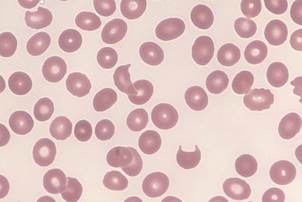 Image S2: bite cells
 Image S3: blister cells