Заглавная страница Избранные статьи Случайная статья Познавательные статьи Новые добавления Обратная связь КАТЕГОРИИ: ТОП 10 на сайте Приготовление дезинфицирующих растворов различной концентрацииТехника нижней прямой подачи мяча. Франко-прусская война (причины и последствия) Организация работы процедурного кабинета Смысловое и механическое запоминание, их место и роль в усвоении знаний Коммуникативные барьеры и пути их преодоления Обработка изделий медицинского назначения многократного применения Образцы текста публицистического стиля Четыре типа изменения баланса Задачи с ответами для Всероссийской олимпиады по праву 
Мы поможем в написании ваших работ! ЗНАЕТЕ ЛИ ВЫ?
Влияние общества на человека
Приготовление дезинфицирующих растворов различной концентрации Практические работы по географии для 6 класса Организация работы процедурного кабинета Изменения в неживой природе осенью Уборка процедурного кабинета Сольфеджио. Все правила по сольфеджио Балочные системы. Определение реакций опор и моментов защемления |
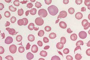
Image S6: irregularly contracted cells
Unstable haemoglobin
J. Burthem, M. Brereton
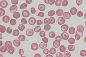
Image S7: schistocytes
Thrombotic thrombocytopenic purpura – a wide range of fragmented red cells with polychromatic cells and other damaged cells. Platelets are absent from the film
J. Burthem, M. Brereton
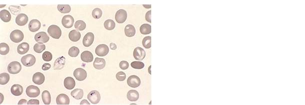
Image S8: sickle cells
Sickle cell disease – typical forms together with other features, notably polychromasia, target cells, and a spherocytic cell.
J. Burthem, M. Brereton 3
Supplementary images: ICSH Recommendations for Peripheral Blood Cell Morphology
Standardization and Grading
Image S9: spherocytes
Autoimmune haemolytic anaemia – typical round, dense red cells
J. Burthem, M. Brereton
Image S10: stomatocytes
Hereditary stomatocytosis
J. Burthem, M. Brereton
Image S11: target cells
Haemoglobin C disease – target cells in association with irregularly contracted cells
J. Burthem, M. Brereton
Image S12: tear drop cells
Myelofibrosis
J. Burthem, M. Brereton 4
Supplementary images: ICSH Recommendations for Peripheral Blood Cell Morphology
Standardization and Grading
Image S13: basophilic stippling
Myelofibrosis - two stippled cells, one in teardrop form
J. Burthem, M. Brereton
Image S14: Howell-Jolly bodies
Auto-splenectomised patient with sickle cell disease
J. Burthem, M. Brereton
Image S15: Pappenheimer bodies
Sideroblastic anaemia – red cells contain small basophilic inclusions of variable size and shape in a limited cytoplasmic area
G. Rozenberg
(Copyright: Microscopic haematology: a practical guide for the laboratory 3e (c) 2011, Sydney, Elsevier Australia)
Image S16: nucleated red blood cell
Myelofibrosis – a typical late stage nucleated red cell in circulation
J. Burthem, M. Brereton 5
Supplementary images: ICSH Recommendations for Peripheral Blood Cell Morphology
Standardization and Grading
Image S17: large granular lymphocyte
Large granular lymphocyte from a normal individual
J. Burthem, M. Brereton
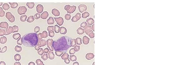
Image S18: Auer rods
AML – two blast cells containing relatively blunt-ended, single and multiple Auer rods
J. Burthem, M. Brereton
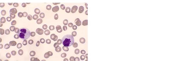
Image S19: hypergranulation
(neutrophils)
Hypergranular neutrophils post G-CSF treatment
J. Burthem, M. Brereton
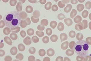
Image S20: hypogranulation
(neutrophils)
Myelodysplasia – hypogranular neutrophils.
Note also the atypical nuclear forms
G. Rozenberg
(Copyright: Microscopic haematology: a practical guide for the laboratory 3e (c) 2011, Sydney, Elsevier Australia) 6
Supplementary images: ICSH Recommendations for Peripheral Blood Cell Morphology
Standardization and Grading
|
||||
|
|
Последнее изменение этой страницы: 2021-03-09; просмотров: 438; Нарушение авторского права страницы; Мы поможем в написании вашей работы! infopedia.su Все материалы представленные на сайте исключительно с целью ознакомления читателями и не преследуют коммерческих целей или нарушение авторских прав. Обратная связь - 3.141.202.54 (0.006 с.) |