Заглавная страница Избранные статьи Случайная статья Познавательные статьи Новые добавления Обратная связь FAQ Написать работу КАТЕГОРИИ: ТОП 10 на сайте Приготовление дезинфицирующих растворов различной концентрацииТехника нижней прямой подачи мяча. Франко-прусская война (причины и последствия) Организация работы процедурного кабинета Смысловое и механическое запоминание, их место и роль в усвоении знаний Коммуникативные барьеры и пути их преодоления Обработка изделий медицинского назначения многократного применения Образцы текста публицистического стиля Четыре типа изменения баланса Задачи с ответами для Всероссийской олимпиады по праву 
Мы поможем в написании ваших работ! ЗНАЕТЕ ЛИ ВЫ?
Влияние общества на человека
Приготовление дезинфицирующих растворов различной концентрации Практические работы по географии для 6 класса Организация работы процедурного кабинета Изменения в неживой природе осенью Уборка процедурного кабинета Сольфеджио. Все правила по сольфеджио Балочные системы. Определение реакций опор и моментов защемления |
Totipotent, multipotent, pluripotent animal cellsСодержание книги Поиск на нашем сайте Totipotent, multipotent, pluripotent animal cells Totipotency is the ability of a single cell to divide and produce all of the differentiated cells in an organism, including extraembryonic tissues.Totipotent cells include spores and zygotes. In the spectrum of cell potency, totipotency represents the cell with the greatest differentiation potential. Toti comes from the Latin totus which means "entirely." Research has helped demonstrate that cells can regain totipotency[5] but research has also shown that regaining totipotency is complex. Instead of regaining full totipotency, cells may instead differentiate through a complex cellular variation of totipotency. Just how totipotent cells arise is described through the process of human development. Consider first that human development begins when a spermfertilizes an egg. The resulting fertilized egg creates a single totipotent cell, a zygote. In the first hours after fertilization, this cell divides into identical totipotent cells, which can later develop into any of the three germ layers of a human (endoderm, mesoderm, or ectoderm) and into cells of thecytotrophoblast layer or syncytiotrophoblast layer of the placenta. After reaching the 16-cell stage, the totipotent cells of the moruladifferentiate into cells that will eventually become either the blastocyst's Inner cell mass or the outer trophoblasts. Approximately four days after fertilization and after several cycles of cell division, these totipotent cells begin to specialize. The inner cell mass, the source ofembryonic stem cells, is pluripotent, not totipotent. In cell biology, pluripotency (from the Latin plurimus, meaning very many, and potens, meaning having power)[11] refers to a stem cell that has the potential to differentiate into any of the three germ layers: endoderm (interior stomach lining, gastrointestinal tract, the lungs), mesoderm (muscle, bone, blood, urogenital), or ectoderm (epidermal tissues and nervous system). Multipotency describes progenitor cells which have the gene activation potential to differentiate into multiple, but limited cell types. For example, a multipotent blood stem cell is a hematopoieticcell — and this cell type can itself differentiate into several types of blood cell types like lymphocytes, monocytes, neutrophils, etc., but cannot differentiate into brain cells, bone cells or other non-blood cell types. New research related to multipotent cells suggests that multipotent cells may be capable of conversion into unrelated cell types. In one case, fibroblasts were converted into functional neurons.[19]In another case, human umbilical cord blood stem cells were converted into human neurons.[21] Research is also focusing on converting multipotent cells into pluripotent cells. Multipotent cells are found in many, but not all human cell types. Multipotent cells have been found in adipose tissue,[23] cardiac cells,[24] bone marrow, and mesenchymal stromal cells (MSCs) which are found in the third molar.
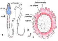
Cryopreservation of reproductive and germ cells of animals and humans Cryopreservation is a process where cells, whole tissues, or any other substances susceptible to damage caused by chemical reactivity or time are preserved by cooling to sub-zero temperatures. At low enough temperatures, any enzymatic or chemical activity which might cause damage to the material in question is effectively stopped. Cryopreservation methods seek to reach low temperatures without causing additional damage caused by the formation of ice during freezing. Traditional cryopreservation has relied on coating the material to be frozen a class of molecules termed cryoprotectants. New methods are constantly being investigated due to the inherent toxicity of many cryoprotectants. Human oocyte cryopreservation (egg freezing) is a novel technology in which a woman’s eggs (oocytes) are extracted, frozen and stored. Later, when she is ready to become pregnant, the eggs can be thawed, fertilized, and transferred to the uterus as embryos. Method The egg retrieval process for oocyte cryopreservation is the same as that for in vitro fertilization. This includes one to several weeks of hormone injections that stimulate ovaries to ripen multiple eggs. When the eggs are mature, a medication to trigger ovulation is given and the eggs are removed from the body using an ultrasound-guided needle through the vagina. The procedure is usually conducted under sedation. The eggs are immediately frozen. The egg is the largest cell in the human body and contains a great amount of water. When the egg is frozen, the ice crystals that form can destroy the integrity of the cell. To prevent this, the egg must be dehydrated prior to freezing. This is done using cryoprotectants which replace the water within the cell and inhibit the formation of ice crystals. Eggs (oocytes) are frozen using either a controlled-rate, slow-cooling method or a newer flash-freezing process known as vitrification. Vitrification is much faster but requires higher concentrations of cryoprotectants to be added. The result of vitrification is a solid glass-like cell, free of ice crystals. Vitrification is associated with higher survival rates and better development compared to slow-cooling when applied to oocytes in metaphase II (MII).[2] Vitrification has also become the method of choice for pronuclear oocytes, although prospective randomized controlled trials are still lacking.[2] Once frozen, the zona pellucida, or shell of the egg hardens. Thus, currently, when eggs are thawed, a special fertilization procedure is performed by an embryologist whereby sperm is injected directly into the egg with a needle rather than allowing sperm to penetrate naturally by placing it around the egg in a dish. This injection technique is called ICSI (Intracytoplasmic Sperm Injection) and is also used in IVF. Semen cryopreservation is a procedure to preserve sperm cells. Semen can be used successfully indefinitely after cryopreservation. For human sperm, the longest reported successful storage is 21 years. It can be used for sperm donation where the recipient wants the treatment in a different time or place, or for men undergoing a vasectomy to still have the option to have children. Freezing The most common cryoprotectant used for semen is glycerol (10% in culture medium). Often sucrose or other di-, trisaccharides are added to glycerol solution. Cryoprotectant media may be supplemented with either egg yolk or soy lecithin, with the two having no statistically significant differences compared to each other regarding motility, morphology, ability to bind to hyaluronate in vitro, or DNA integrity after thawing. [1] Semen is frozen using either a controlled-rate, slow-cooling method (slow programmable freezing or SPF) or a newer flash-freezing process known as vitrification. Vitrification gives superior post-thaw motility and cryosurvival than slow programmable freezing. [2] Thawing Thawing at 40°C seems to result in optimal sperm motility. On the other hand, the exact thawing temperature seems to have only minor effect on sperm viability, acrosomal status, ATP content, and DNA. Refreezing In terms of the level of sperm DNA fragmentation, up to three cycles of freezing and thawing can be performed without causing a level of risk significantly higher than following a single cycle of freezing and thawing. This is provided that samples are refrozen in their original cryoprotectant and are not going through sperm washing or other alteration in between, and provided that they are separated by density gradient centrifugation or swim-up before use in assisted reproduction technology. Techniques Culture of embryos can either be performed in an artificial culture medium or in an autologous endometrial coculture (on top of a layer of cells from the woman's own uterine lining). With artificial culture medium, there can either be the same culture medium throughout the period, or a sequential system can be used, in which the embryo is sequentially placed in different media. For example, when culturing to the blastocyst stage, one medium may be used for culture to day 3, and a second medium is used for culture thereafter.[2] Single or sequential medium are equally effective for the culture of human embryos to the blastocyst stage.[3] Artificial embryo culture media basically contain glucose, pyruvate, and energy-providing components, but the addition of amino acids, nucleotides, vitamins, and cholesterol improve the performance of embryonic growth and development.[4] Methods to permit dynamic embryo culture with fluid flow and embryo movement are also available.[5] A new method in development uses the uterus as an incubator and the naturally occurring intrauterine fluids as culture medium by encapsulating the embryos in permeable intrauterine vessel. Size and shape The egg cell (or ovum, or oocyte) is the largest human cell. She measures 0.15 to 0.2 mm and is just visible to the naked eye. She is also the roundest cell, she is almost perfectly round (Fig. 4). She therefore has the largest volume in relation to her surface. The cell consists of a large amount of cytoplasm (= cell fluid) in which the nucleus is dissolved (and therefore invisible) until just before conception. Sperm cells are the smallest human cells. They are no more than a nucleus with a small amount of cytoplasm, some mitochondria (the energy suppliers of the cell) and a long tail. They have hardly any content and are the straightest cells.
Mobility In contrast, the ovum is externally not active. After her release, she is passively moved by the fluid-flow in the oviduct (uterine tube), while the sperm cells are active, using their tails to swim against the stream of fluid in the oviduct. They are externally active and mobile. The ovum is internally mobile and externally passive, this is a polarity. The sperm shows the opposite: internally passive and externally mobile. Egg cell and sperm have a polarity and are opposite to each other, we see a double polarity. Sperm cells do not absorb or release substances. There is no interaction with the environment. They live about 3 to 5 days in the womb and can be preserved and frozen at temperatures below 60 °C. They are not easy to destroy. They are closed off from the environment and metabolically passive. The open and vulnerable state of the egg cell is polar to the closed and robust state of the sperm cells. The ovum is alone and the sperm are with millions. One sperm cell is nothing, one ovum determines everything. One is polar to millions. One comprises everything, it is all there is, whereas the millions of sperm cells are infinitive, have no importance on their own. Location The ovum develops in warm- and sperm in relative cold conditions. Development Egg cells are old cells that became mature. Primordial oocytes are in a process of dying. Sperm cells are newly formed and are young. The maturation process of ova is an expiring process, it stops. The formation of the sperm is a vital process, it never stops. 39) Basic approaches and principles of gene therapy. Gene therapy ex vivo, in vivo, in situ. Gene therapy is the use of DNA as a pharmaceutical agent to treat disease. It derives its name from the idea that DNA can be used to supplement or alter genes within an individual's cells as a therapy to treat disease. The most common form of gene therapy involves using DNA that encodes a functional, therapeutic gene to replace a mutated gene. Other forms involve directly correcting a mutation, or using DNA that encodes a therapeutic protein drug (rather than a natural human gene) to provide treatment. In gene therapy, DNA that encodes a therapeutic protein is packaged within a "vector", which is used to get the DNA inside cells within the body. Once inside, the DNA becomes expressed by the cell machinery, resulting in the production of therapeutic protein, which in turn treats the patient's disease. Gene therapy may be classified into the two following types: [edit] Somatic gene therapy In somatic gene therapy, the therapeutic genes are transferred into the somatic cells, or body, of a patient. Any modifications and effects will be restricted to the individual patient only, and will not be inherited by the patient's offspring or later generations. Somatic gene therapy represents the mainstream line of current basic and clinical research, where the therapeutic DNA transgene (either integrated in the genome or as an external episome or plasmid) is used to treat a disease in an individual. [edit] Germ line gene therapy In germ line gene therapy, germ cells, i.e., sperm or eggs, are modified by the introduction of functional genes, which are integrated into their genomes. This would allow the therapy to be heritable and passed on to later generations. Although this should, in theory, be highly effective in counteracting genetic disorders and hereditary diseases, many jurisdictions prohibit this for application in human beings, at least for the present, for a variety of technical and ethical reasons This can be accomplished in three ways: ex vivo, in situ, or in vivo. Ex vivo involves removing cells from the patient, altering the genetic material, and placing them back into the patient. In situ requires the vector be placed directly into the affected tissues. In vivo gene therapy involves injecting the vector into the bloodstream. Draw a diagram of an experiment in genetic engineering (design recDNA) and give a description of the main stages Molecular cloning is the laboratory process used to create recombinant DNA. It is one of two widely used methods (along withpolymerase chain reaction, abbr. PCR) used to direct the replication of any specific DNA sequence chosen by the experimentalist. The fundamental difference between the two methods is that molecular cloning involves replication of the DNA within a living cell, while PCR replicates DNA in the test tube, free of living cells. Formation of recombinant DNA requires a cloning vector, a DNA molecule that will replicate within a living cell. Vectors are generally derived from plasmids or viruses, and represent relatively small segments of DNA that contain necessary genetic signals for replication, as well as additional elements for convenience in inserting foreign DNA, identifying cells that contain recombinant DNA, and, where appropriate, expressing the foreign DNA. The choice of vector for molecular cloning depends on the choice of host organism, the size of the DNA to be cloned, and whether and how the foreign DNA is to be expressed.[5] The DNA segments can be combined by using a variety of methods, such as restriction enzyme/ligase cloning or Gibson assembly. In standard cloning protocols, the cloning of any DNA fragment essentially involves seven steps: (1) Choice of host organism and cloning vector, (2) Preparation of vector DNA, (3) Preparation of DNA to be cloned, (4) Creation of recombinant DNA, (5) Introduction of recombinant DNA into the host organism, (6) Selection of organisms containing recombinant DNA, (7) Screening for clones with desired DNA inserts and biological properties.
Construction of recombinant DNA, in which a foreign DNA fragment is inserted into a plasmid vector. In this example, the gene indicated by the white color is inactivated upon insertion of the foreign DNA fragment. GIFT (gamete intrafallopian transfer) and ZIFT (zygote intrafallopian transfer) are modified versions of in vitro fertilization. Like IVF, these procedures involve retrieving an egg from the woman and re-implanting it after manipulation. Unlike IVF, the timing between mixing the sperm and eggs and the transfer is faster. While the success rates are similar to IVF, the processes used in GIFT and ZIFT are closer to natural conception. In ZIFT, the eggs are placed in the fallopian tubes rather than directly in the uterus. With GIFT, fertilization actually takes place in the body rather than in a petri dish. As in vitro fertilization techniques have become more refined, GIFT and ZIFT have become less relied upon. Additionally, as GIFT and ZIFT require surgery while IVF does not, IVF is the preferred choice in clinics. In vitro fertilization accounts for at least 98% of all assisted reproductive technology procedures performed in the U.S., while GIFT and ZIFT make up less than 2%. What Types of Infertility Are Addressed By Gift and Zift? GIFT and ZIFT can be used to treat many types of infertility, except cases caused by damage or abnormalities of the fallopian tubes. These techniques can also be used in cases of mild male infertility, as long as the sperm is capable of fertilizing an egg. GIFT: What You Can Expect Eggs and sperm are collected just as they would be in an IVF procedure, but after that, the two techniques differ. Unlike IVF, GIFT requires an incision be made in the abdomen and the eggs and sperm placed in the fallopian tubes using a laparoscope. Because the eggs and sperm are placed into the fallopian tubes before conception, there's no way to know if fertilization has taken place prior to transfer. Typically, more eggs will be used in GIFT to ensure pregnancy, which has the side effect of increased multiple births. ZIFT: What You can Expect Unlike GIFT, with ZIFT the sperm and egg are mixed together in the laboratory, and given time to fertilize before being transferred to the fallopian tubes thus lowering the number of eggs used and correspondingly the chances for multiple pregnancy. As this is a post-fertilization technique, ZIFT is closer to in vitro fertilization. ZIFT, like GIFT, requires the procedure be performed by laparoscopy.
Totipotent, multipotent, pluripotent animal cells Totipotency is the ability of a single cell to divide and produce all of the differentiated cells in an organism, including extraembryonic tissues.Totipotent cells include spores and zygotes. In the spectrum of cell potency, totipotency represents the cell with the greatest differentiation potential. Toti comes from the Latin totus which means "entirely." Research has helped demonstrate that cells can regain totipotency[5] but research has also shown that regaining totipotency is complex. Instead of regaining full totipotency, cells may instead differentiate through a complex cellular variation of totipotency. Just how totipotent cells arise is described through the process of human development. Consider first that human development begins when a spermfertilizes an egg. The resulting fertilized egg creates a single totipotent cell, a zygote. In the first hours after fertilization, this cell divides into identical totipotent cells, which can later develop into any of the three germ layers of a human (endoderm, mesoderm, or ectoderm) and into cells of thecytotrophoblast layer or syncytiotrophoblast layer of the placenta. After reaching the 16-cell stage, the totipotent cells of the moruladifferentiate into cells that will eventually become either the blastocyst's Inner cell mass or the outer trophoblasts. Approximately four days after fertilization and after several cycles of cell division, these totipotent cells begin to specialize. The inner cell mass, the source ofembryonic stem cells, is pluripotent, not totipotent. In cell biology, pluripotency (from the Latin plurimus, meaning very many, and potens, meaning having power)[11] refers to a stem cell that has the potential to differentiate into any of the three germ layers: endoderm (interior stomach lining, gastrointestinal tract, the lungs), mesoderm (muscle, bone, blood, urogenital), or ectoderm (epidermal tissues and nervous system). Multipotency describes progenitor cells which have the gene activation potential to differentiate into multiple, but limited cell types. For example, a multipotent blood stem cell is a hematopoieticcell — and this cell type can itself differentiate into several types of blood cell types like lymphocytes, monocytes, neutrophils, etc., but cannot differentiate into brain cells, bone cells or other non-blood cell types. New research related to multipotent cells suggests that multipotent cells may be capable of conversion into unrelated cell types. In one case, fibroblasts were converted into functional neurons.[19]In another case, human umbilical cord blood stem cells were converted into human neurons.[21] Research is also focusing on converting multipotent cells into pluripotent cells. Multipotent cells are found in many, but not all human cell types. Multipotent cells have been found in adipose tissue,[23] cardiac cells,[24] bone marrow, and mesenchymal stromal cells (MSCs) which are found in the third molar.
|
||
|
|
Последнее изменение этой страницы: 2016-08-06; просмотров: 274; Нарушение авторского права страницы; Мы поможем в написании вашей работы! infopedia.su Все материалы представленные на сайте исключительно с целью ознакомления читателями и не преследуют коммерческих целей или нарушение авторских прав. Обратная связь - 216.73.216.214 (0.011 с.) |