Заглавная страница Избранные статьи Случайная статья Познавательные статьи Новые добавления Обратная связь КАТЕГОРИИ: ТОП 10 на сайте Приготовление дезинфицирующих растворов различной концентрацииТехника нижней прямой подачи мяча. Франко-прусская война (причины и последствия) Организация работы процедурного кабинета Смысловое и механическое запоминание, их место и роль в усвоении знаний Коммуникативные барьеры и пути их преодоления Обработка изделий медицинского назначения многократного применения Образцы текста публицистического стиля Четыре типа изменения баланса Задачи с ответами для Всероссийской олимпиады по праву 
Мы поможем в написании ваших работ! ЗНАЕТЕ ЛИ ВЫ?
Влияние общества на человека
Приготовление дезинфицирующих растворов различной концентрации Практические работы по географии для 6 класса Организация работы процедурного кабинета Изменения в неживой природе осенью Уборка процедурного кабинета Сольфеджио. Все правила по сольфеджио Балочные системы. Определение реакций опор и моментов защемления |
The topic studied actuality.
Doctor often meets excitement conductance disorders in nervous-muscular synapses and nervous fibers. He must influence on them well-singlemindedly with impulses conductance enforcement or weakening. So, he should know excitement conductance mechanisms and regularities. Nervous fiber is reflectory arc afferent or efferent part. Reflex disappears at nervous impulse conductance disturbance. Different nervous fibers possess various excitability and excitement conductance different speed. It should be taken into account while medicines dosage prescription. Nervous fibers conductance laws are used in neurology and anaesthesiology. Axons can be injured at some diseases. Both distal and proximal part can degenerate after axon cutting. It regenerates in peripheral nervous system after several weeks but its growing locuses first are non-myelinized. At different-origined neuropathies axons also loose their myeline sheath becoming demyelinated. Besides, doctor can meet axone neuropathies main symptome of which is, probably, axon transport disorder. Demyelinated axons especially often interact with abnormal interactions. Impulses passing through nervous fibers groups induce the excitement of other parallel axons. So, law of impulse isolated coming (see below) becomes injured. It is known as ephaptic transmission. When such abnormal action potentials are generated in sensory nervous fibers abnormal sensations appear in patient. They can be agonizing especially if they are connected with nociceptive (pain) fibers. Neuralgy (pain by nervous fibers course), causalgy (pain without definite origine), neuromic pain and other hardly-treated and non-pleasant syndromes can appear. Interaxonal problems can be the result both of insufficient isolation (with myeline sheathes) and increased axons excitability. Multiple sclerosis is a neurological disease usually diagnosed in people between the ages of 20 and 40. It is a chronic, degenerating, remitting, and relapsing disease that progressively destroys the myelin sheaths of neurons in multiple areas of the CNS. Initially, lesions form on the myelin sheaths and soon develop into hardened scleroses, or scars (from the Greek word sclerosis, meaninf “hardened”). Destruction of the myelin sheaths prohibits the normal conduction of impulses, resulting in a progressive loss of functions. Because myelin degeneration is widespread and affects different areas of the nervous system in different people, multiple sclerosis has a wider variety of symptoms than any other neurological disease. Although the causes of it are not fully known, there is evidence that the disease involves a genetic susceptibility combined with an immune attack on the oligodendrogliocytes and myelin, perhaps triggered by viruses. Inflammation and demyelination then occur, leading to the symptoms of multiple sclerosis. Alongside with exciting or inhibitory functional signals transmittance, synapses provide trophic (dealing with growth and differentiation) interconnections of contacting cells realized by means of trophic proteinic factors (probably accumulating in “dark” vesicles). Such influencings are especially expressed in neuro-muscular (myoneural) synapses directed to myocytes metabolic support. Denervation leads to skeletal muscular fibers features changing: to fast muscles retardation as well as slow ones acceleration id est to differentiation loosing. Muscular denervation effects are imitated with actions of toxins which disturb synaptic transmission namely botulinic and diphtherial one. They know high synapses sensitivity to different influencing which makes these neuronal parts the mostly vulnerable. Moreover, switching definite synapses quantity off brain function and injury of any synapses system are able to form clinical symptomal complexes by themselves, influence on brain diseases dynamics as a whole. Electronic microscopy can show several clinical syndromes which are developed only or first of all due to synapses injuries.
Synapses ultrastructure pathological changings are not specific to aethiological moments caused their injury. Synapses injuries reasons: 1. ionizing irradiation; 2. electrical trauma; 3. pharmacological actions; 4. intoxications; 5. hypoxy. It makes tryings to classify synapses pathological changings by traditional ethiological principle rather difficult. Absence of rather detailed information about local physiological and biochemical changings in synapses does not allow apply also pathogenetical principle at this classification processing. Moreover, identical by pathogenesis influencings (hypoxy, axon cutting) cause sometimes non-equal changings of axons presynaptic parts even in one system of fibers. 4 main types of synapses injury: 1. dark degeneration (cytoplasm staining with black osmium increasing); 2. filamentary degeneration (like ring); 3. light degeneration (cytoplasm staining with black osmium decreasing); 4. focal degeneration (synapse decomposition). Dark degeneration is the most widely-spread; focal one – the least one. Dark degeneration is observed at axon cutting. Synapses changings by light type occur not only as a result of axons injury but they are characteristic symptom of brain injury at hypoxy. Synapse focal or partial destruction is characterized by non-equal changings of presynapse different parts. Synapses changings at hypoxy: 1.presynaptic process strong swelling; 2.synaptic vesicles amount significant decreasing up to their complete disappearance; 3.synaptic vesicles agglutination (gluing) in groups located near presynaptic membrane in axon center; 4.mitochondria decomposition; 5.appearance other structures absent under normal conditions in presynaptic processes such as lysosomes, large vacuoles, concentric membranes accumulations; 6.synapses partial focal destruction which is of a special significance for tiny pathological mechanisms understanding. Local anesthesia physiological basement in dentistry. Significance of excitement conductance laws (through nervous fibers) in dentistry. Nervous fibre physiological integrity law is used in dental practice for local anesthesia performance: nervous impulse conductance through nervous fibre is possible only at its physiological integrity; narcotic substance, cooling action, ligature or weak electrical current influence lead to nervous fibre structures functioning stoppage and prevent excitement distribution. Conductive anaesthesia - analgetic (novocaine) introduction - and excitement in pharmacological blockade zone is not distributed. Electroanalgesia – anode (positive electrode) putting from current origin blockates receptory cells membranes depolarization; that is why noceoceptive impulses do not occur. Electrical magnetical waves application – analgesia is the result of neurons membranes depolarization prevention. Non-medical analgesia methods (cooling, electroanalgesia, reflexoanalgesia) physiological basement. Possibilities and prospects of their usage in dentistry. Cooling in dentistry is applied for superficial local anaesthesia. It is developed because oral mucosa temperature decreasing leads to ischemia, sensitivity inhibiting (nervous formations excitability decreasing). Electroanalgesia – see above. Reflexoanalgesia – is based on reflectory action to the sick organ by peripheral biologically-active points irritation. Analgetic effects appears by means of somatic and visceral signals convergence on neurons of CNS different levels. At polymodal signals convergence somatic impulses are dominant over visceral ones. That is why impulses flow from skin-muscular nerves if it precedes to visceral one will inhibit it. Besides, the same relations are possible also inside somatic nervous system between impulsation on fast-conducting fibers which transmit dull, whining pain; impulses on fast fibers inhibits entrance on spinal neurons while this and the reactions to following stimuli on slow fibers are blocked.
2.Study aims: To know: excitement developmental mechanisms in myeline and myeline-free nervous fibers; main factors determining conductance velocity; synapses main types; excitement regularities in chemical and electrical synapses. To be able to: interpret conductivity disorders reasons, excitement nervous-muscular conductance blockade mechanisms; to draw schematically conductance mechanism in myelin and myelin-free fibers as well as through nervous-muscular synapse.
3.Pre-auditory self-work materials. 3.1.Basic knowledge, skills, experiences, necessary for study the topic:
Topic content. Structural and functional unit of nervous system and nervous tissue is defined as neuron or nerve cell. Neuronal organization main principles (by R.Kahal): 1. Neuron with its processes represents one morphological whole. 2. Neurons are similar genetically, they are origined from one neuroblasts. 3. Neuron is one functional integrity. 4. Excitement alongside neuron is distributed in 1 direction – from dendrites to axon. 5. Neuron represents trophyc integrity. Axons death is observed at neurons bodies removal. Axon cutting also leads to neurons death. 6. Neuron participates in pathological reactions as one a whole. Destructive changings engulf a whole neuron. Neuronal theory. Mentioned principles are in the basement of CNS structure neuronal theory. According to this theory, CNS structure and function are defined by multiplied interconnected neurons with discrete features. Also CNS represents continuous structure like syncitium (it represents more in CNS of Non-Vetebrata (Hydras and other Coelenterata) and Amphibias. Heterogenous neurons in their interconnections allow human CNS participate in millions various reactions. From hundreds to thousands different synapses are located on separate neurons bodies. Separate synapses function with different mediators and are connected to specific postsynaptic chemical reactions (see below). Excitement process in neurons can be explained by electric and chemical theory. Electrical theory. Morphological-functional neuronal variery lies on its base. There is so-called “light axonic colliculus” or “light spot” at the place of axons divergence. This neuron locus is charged positively comparatively to neurons membranes other locusi (they have negative charge). At excitement of synapses located on neurons dendrite and body potentials difference is increased between “axons colliculus” and rest neuron part. It gives life to the axon spike activity at definite level reaching. The more is gradient of potentials difference between neuron body and “axon colliculus”, the more frequent is initial axonic impulsation. Chemical theory This theory connects neurons excitement processes with specificity of mediators, oligopeptides and ther biologically-active substances released from synapses. You will study below about excitable (noradrenaline, acetylcholine, glutamate) and inhibitory (gamma-aminooleic acid, glycine) mediators in synapses. Also neurons excitement process depends on the specificity of postsynaptic chemical reactions which are distributed to the neurons nuclei genetic apparatus.
NEURONS CLASSIFICATION 1. Depending on number of poles: a) unipolar neurons – are present only in embryonic stage in human beings, from the single pole, both the processes - axon and dendrite arise; b) pseudounipolar – they have one axon and one dendrite but both come from one neuron body pole; example – spinal ganglii; c) bipolar neurons - have two poles, one for axon and another for dendrite; example – retina, cochlea spiral ganglium; d) multipolar neurons - have many poles, one of which gives rise to the axon and all the other poles give rise to dendrites; biggest neurons number belong to this group.
2. Depending on function: a) motor or efferent neurons - carry the motor impulses from central nervous system to the peripheral effector organs like muscles, glands, blood vessels, etc.: § neurons of autonomic nervous system; § alha-motoneurons – innervate extrafusal muscular fibers (out of muscular spindles); § gamma-motoneurons – innervate intrafusal muscular fibers (inside muscular spindles); b) sensory or afferent neurons - neurons carry the sensory impulses from periphery to the central nervous system; c)associative (99,8% of all) – they bind different-typed neurons. P.S. Muscular spindles – are muscles receptors percepting muscle length and its contraction speed.
3. Depending on length of axon: a) Golgi Type I Neurons - have long axons, the cell body of these neurons is in central nervous system and their axons reach remote (far) peripheral organs; b) Golgi Type II Neurons - have short axons, are present in cerebral cortex and spinal cord, cerebellar Purkin’e cells belong to them.
4. Depending on mediator: a) adrenergic – contains noradrenalin; b) cholinergic – acetylcholine; c) dopaminergic – neuromediator dopamine; d) peptidergic – contain one of neuropeptides: § substance P; § neuropeptide Y; § calcitonin-gene-related peptide and so on. NEURONS STRUCTURE 1. Nerve cell body 2. Dendrite 3. Axon 4. Myelin sheath or membrane 5. Neurilemma Dendrites are conductive in nature and transmit impulses towards the nerve cell body. Dendrites are shorter processes terminating mostly near nerve cell body. Axon is longer process of nerve cell. Axon may extend for a long distance away from nerve cell body. Length of the longest axon is about one meter. NEUROGLIA It is located between neurons. These cells, gliocytes, are known as supporting cells. The name origin: Greek – “neuron” – “nerve” and “glia” – “glue”. Functions: 1. supporting; 2. trophic; 3. demarcating; 4. barrier; 5. secretory; 6. protective. 2 MAIN TYPES: 1. Microglia – microgliocytes (main CNS phagocytes). 2. Macroglia: § astrocytes; § ependimocytes; § oligodendrogliocytes. Astrocytes are the biggest gliocytes. Protoplasmic astrocytes are located in grey matter; fibrous astrocytes are situated in white matter. Functions: 1. supporting – CNS carcass forming especially during embryogenesis; directing – synthesis of substances determining neurons differentiation; 2. demarcating, transport and barrier – is directed to neurons optimal microenvironment providing: a) hemato-encephalic barrier (HEB) – it separates CNS neurons from blood and internal environment tissues and includes: – capillaries endothelium; – basal membrane; – perivascular membrane (near-vessel) formed by astrocytes processuses; b) neuro-liquor barrier – it separates neurons from liquor; c) formation of perineuronal sheathes surrounding neurons bodies in synaptic area (isolating function); 3. metabolic and regulatory – one of the most important astrocytes functions – K+-ions and mediators optimal level supporting in neurons surrounding; 4. protective: – phagocytic; – immune (antigen-presenting cells); – reparative (they form scar at ending stages of inflammation in CNS while their growth).
Ependimocytes – (“ependyma” – “street cloth” or “covering”) cover brain ventricles and cerebral sheathes. Functions: 1. supporting; 2. forming of barriers: – neural-liquor (with high permeability); – hemato-liquor; 3. liquor components ultrafiltration. Tanicites – cover the III-rd ventricle walls. They absorb substances from liquor and transport them through their process to vessels lumen thus providing connection between liquor in ventricles lumen and blood. Oligodendrogliocytes – (Greek – “oligo” – “a little”, “dendron” – “tree” and “glia” – “glue” - glia with little processuses number – they surround neurons bodies, are in nervous endings compounds; they are observed both in central and peripheral nervous system. Functions: 1. myeline sheath formation; 2. neurons metabolism.
Neurotrophins In a developing fetal brain, chemicals called neurotrophins, promote neuron growth. Nerve growth factor (NGF) was the first neurotrophin to be identified; others include brain-derived neurotrophic factor (BDNF); glial-derived neurotrophic factor (GDNF); neurotrophin-3 and neurotrophin 4/5 (the numbers depend on the species). NGF and neutrophin-3 are known to be particularly important in the embryonic development of sensory neurons and sympathetic ganglia. Neurotrophins also have important functions in the adult nervous system. NGF is required for the maintenance of sympathetic ganglia, an there is evidence that neurotrophins are required for mature sensory neurons to regenerate after injury. In addition, GDNF may be needed in the adult to maintain spinal motor neurons and to sustain neurons in the brain that use the chemical dopamine as a neurotransmitter. Experiments suggest that neurons of the CNS can regenerate if they are provided with the appropriate environment. While neutrophins promote neuron growth, some chemicals including myelin-associated inhibitory proteins, have bben shown to inhibit axon regeneration. Research in this area, with its important implications for the repair of spinal cord and brain injury, is ongoing.
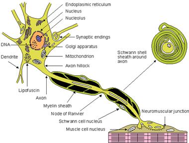
NERVOUS FIBERS PHYSIOLOGY Structure of Myelinated Nerve Fiber Axis cylinder of nerve fiber is covered by a membrane called neurilemma. In myelinated nerve fiber, axis cylinder is covered by a thick sheath called myelin sheath. Myelin sheath in turn is covered by neurilemma.
Fig.10. Myelinated nerve fiber Myelin sheath In a myelinated nerve fiber, axis cylinder is covered by a thick tubular sheath called myelin sheath. Myelin sheath does not form a continuous sheath and is absent at regular intervals. The area where the myelin sheath is absent is called node of Ranvier. The segment of nerve fiber between two nodes is called internode. Myelin sheath is responsible for white colour of nerve fibers. Myelin is lipoproteid. Functions of Myelin Sheath Myelin sheath is responsible for faster conduction of impulse through nerve fibers. In these nerve fibers, impulses jump from one node to another. Transmission of impulses from one node to another is by means of saltatory conduction. Myelin sheath also has a high insulating capacity. Because of this, myelin sheath restricts nerve impulse within single nerve fiber, and prevents stimulation of neighboring nerve fibers. Neurilemma Surrounding myelin sheath, there is a thin membrane called neurilemmal sheath. This is also called neurilemma or sheath of Schwann. It contains Schwann cells. Cytoplasm is thin and modified to form the thin sheath of neurilemma enclosing the myelin sheath. At node of Ranvier (where myelin sheath is absent), the neurilemma invaginates and runs up to axolemma in form of a finger-like process. Neurilemma is necessary for formation of myelin sheath (myelinogenesis). Neurilemma is absent in central nervous system. In non-myelinated nerve fiber, the neurilemma continuously surrounds axolemma.
Nervous impulses conduction
Fig.11. Mode of conduction through nerve fibers. A. Non-myelinated nerve fiber — Continuous conduction. B. Myelinated nerve fiber — Saltatory conduction: impulse jumps from node to node. AP = action potential.
CONDUCTION THROUGH MYELINATED NERVE FIBER—SALTATORY CONDUCTION Conduction of impulse through a myelinated nerve fiber is about 150 times faster than through a non-myelinated fiber. This is because, myelin sheath forms an effective insulator and flow of current through this sheath is negligible. But action potential jumps from one node to another node of Ranvier. So, velocity of conduction is faster. This type of jumping of action potential from one node to another is called saltatory conduction.
|
|||||||||||||||||||||||
|
|
Последнее изменение этой страницы: 2021-03-09; просмотров: 82; Нарушение авторского права страницы; Мы поможем в написании вашей работы! infopedia.su Все материалы представленные на сайте исключительно с целью ознакомления читателями и не преследуют коммерческих целей или нарушение авторских прав. Обратная связь - 3.138.174.195 (0.101 с.) |