Заглавная страница Избранные статьи Случайная статья Познавательные статьи Новые добавления Обратная связь FAQ Написать работу КАТЕГОРИИ: ТОП 10 на сайте Приготовление дезинфицирующих растворов различной концентрацииТехника нижней прямой подачи мяча. Франко-прусская война (причины и последствия) Организация работы процедурного кабинета Смысловое и механическое запоминание, их место и роль в усвоении знаний Коммуникативные барьеры и пути их преодоления Обработка изделий медицинского назначения многократного применения Образцы текста публицистического стиля Четыре типа изменения баланса Задачи с ответами для Всероссийской олимпиады по праву 
Мы поможем в написании ваших работ! ЗНАЕТЕ ЛИ ВЫ?
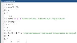
Влияние общества на человека
Приготовление дезинфицирующих растворов различной концентрации Практические работы по географии для 6 класса Организация работы процедурного кабинета Изменения в неживой природе осенью Уборка процедурного кабинета Сольфеджио. Все правила по сольфеджио Балочные системы. Определение реакций опор и моментов защемления |
Vestibulocochlear nerve (CN VIII)Содержание книги Поиск на нашем сайте
Cranial Nerves
OLFACTORY NERVE (CN I) OPTIC NERVE (CN II) OCULOMOTOR NERVE (CN III) TROCHLEAR NERVE (CN IV) TRIGEMINAL NERVE (CN V) ABDUCENT NERVE (CN VI) FACIAL NERVE (CN VII) VESTIBULOCOCHLEAR NERVE (CN VIII) GLOSSOPHARYNGEAL NERVE (CN IX) VAGUS NERVE (CN X) SPINAL ACCESSORY NERVE (CN XI) HYPOGLOSSAL NERVE (CN XII) SYNONYMS AND EPONYMS FOLLOW-UP TO CLINICAL CASE QUESTIONS TO PONDER There are 12 pairs of cranial nerves emerging from the brain and radiating from its surface (Fig. 15.1). They pass through skull foramina, fissures, or canals to exit the cranial vault and then distribute their innervation to their respective structures in the head and neck. One of the cranial nerves, the vagus (L., “wanderer”) continues into the trunk where it innervates various thoracic and abdominal organs. In addition to being named, the cranial nerves are numbered sequentially with Roman numerals in the order in which they arise from the brain, rostrally to caudally. The following list includes their names and corresponding numbers.
Figure 1 = Ventral view of the brainstem showing the cranial nerves Although the cranial nerves and their sensory and parasympathetic ganglia (Tables 15.1, 15.2) form part of the peripheral nervous system, the optic nerve is really an outgrowth of the brain that emerges from the prosencephalon (not the brainstem as other cranial nerves), and is therefore not a typical cranial nerve. Moreover, part of the spinal accessory nerve arises from the cervical spinal cord; thus there are only nine pairs of cranial nerves that emerge from the brainstem. The main sensory and motor nuclei of the cranial nerves are shown in Fig. 15.2. In describing the various functional components (modalities) of the cranial nerves, the definition of the following terms should be kept in mind: afferent is sensory input; efferent is motor output that may be somatic to skeletal muscles or visceral to smooth muscle, cardiac muscle, and glands, and special visceral efferent to striated muscles derived from the brachial arches; general refers to those components that may be carried by cranial nerves as well as spinal nerves; special refers to functional components that are carried by cranial nerves only. The following categories describe the functional components carried by the various cranial nerves (Table 3).
Table.3 = Cranial nerve functional components.
1 General somatic afferent (GSA). These fibers carry general sensation (touch, pressure, pain, and temperature) from cutaneous structures and mucous membranes of the head, and general proprioception (GP) from somatic structures such as muscles, tendons, and joints of the head and neck. The trigeminal, facial, glossopharyngeal, and vagus nerves transmit GSA input to the spinal nucleus of the trigeminal nerve.
2 General somatic efferent (GSE). These fibers provide general motor innervation to skeletal muscles derived from embryonic somites. The oculomotor, trochlear, and abducent nerves innervate the extraocular muscles that Mesencephalic tract and nucleus of CN V Edinger–Westphal nucleus Oculomotor nucleus Trochlear nucleus Motor nucleus of CN V Abducens nucleus. Superior salivatory nucleus Facial nucleus Inferior salivatory nucleus Hypoglossal nucleus Dorsal motor nucleus of CN X Nucleus ambiguous Spinal accessory nucleus Main sensory nucleus of CN V Spinal nucleus of CN V Nucleus of the solitary tract. Figure 15.2 = The nuclei of the cranial nerves. The sensory nuclei are illustrated on the left, and the motor nuclei on the right. OLFACTORY NERVE (CN I) The bipolar olfactory receptor cells (first ordersensory neurons) of theolfactory apparatus residenot in a sensory ganglion,but instead in the olfactory epithelium (neuroepithelium) of the modified nasal mucosa lining the roof and adjacent upper walls of the nasal cavities (see Fig. 19.1).
The axons of these bipolar neurons are SVA fibers transmitting olfactory sensation. These axons assemble to form bundles, the olfactory fila (L., “threads”), which collectively form cranial nerve I. Theolfactory fila traverse the fenestrations of the cribriform plateof the ethmoid bone to terminate in the olfactory bulb wherethey synapse with second order relay neurons and interneurons. OPTIC NERVE (CN II) The optic nerve mediates the special sense of vision via its SSA fibers. Light entering the eye activates cells known as rods and cones, the photoreceptors of the retina. Electrical signals generated by the photoreceptors are transmitted to other cells of the retina that process and integrate sensory input. The first order sensory bipolar neurons of the visual pathway reside in the retina and transmit electrical signals of visual sensory input to the multipolar second order ganglion cells of the retina. The ganglion cells give rise to unmyelinated axons that converge at the optic disc and traverse the lamina cribrosa, a sieve-like perforated area of the sclera, to emerge from the back of the eyebulb. At this point, the ganglion cell axons acquire a myelin sheath and assemble to form the optic nerve. This nerve, an outgrowth of the diencephalon, leaves the orbit via the optic canal to enter the middle cranial fossa.
There, the optic nerves of the right and left sides join each other to form the optic chiasma (G., “optic crossing”) where partial decussation of the optic nerve fibers of the two sides takes place. All ganglion cell axons arising from the nasal half of the retina decussate (through the central region of the chiasma) to the opposite optic tract. All ganglion cell axons arising from the temporal half of the retina proceed (through the lateral aspect of the chiasma) without decussating and join the optic tract of the same side. The ganglion cell axons coursing in each optic tract curve around the cerebral peduncle to terminate and relay visual input in one of the following four regions of the brain: the lateral geniculate nucleus, a thalamic relay station for vision; the superior colliculus, a mesencephalic relay station for vision associated with somatic reflexes; the pretectal area, a mesencephalic region associated with autonomic reflexes; and the hypothalamus (see Figs 16.5, 16.7, 16.9).
OCULOMOTOR NERVE (CN III) The oculomotor nerve supplies skeletal motor (somatomotor) innervationto the superior rectus, medialrectus, inferior rectus, andinferior oblique muscles(which move the bulb of theeye) and the levator palpebrae superioris muscle (whichelevates the upper eyelid). It also provides parasympathetic (visceromotor) innervation to the ciliary and sphincter pupillaemuscles, two intrinsic smooth muscles of the eye.
The triangular-shaped oculomotor nuclear complex islocated in the mesencephalon. It is situated ventral to theperiaqueductal gray, adjacent to the midline at the levelof the superior colliculus. The oculomotor nucleus consistsof several subnuclei representing each of the extraocularmuscles. These subnuclei are composed of groups of nervecell bodies of the GSE neurons that innervate the listedextraocular muscles and the levator palpebrae superiorismuscle. The cell group innervating the levator palpebraesuperioris is located in the midline, sending motor fibers tothis muscle bilaterally (both right and left upper eyelids). The cell group innervating the superior rectus sends projections to the opposite side; whereas the cell group innervating the medial rectus, inferior oblique, and inferior rectus sends projections to the same side. The Edinger–Westphal nucleus, a subnucleus of the oculomotor nuclear complex is located dorsally, medially, and rostral to the GSE nuclear complex. It contains the cell bodies of GVEpreganglionic parasympathetic neurons whose axons join the GSE fibers as they converge and pass ventrally in the midbrain to emerge from the ventral aspect of the brainstem in the interpeduncular fossa as the oculomotor nerve. The oculomotor nerve proceeds anteriorly within the cranial vault, travels within the cavernous sinus, and by passing through the superior orbital fissure, enters the ipsilateral orbit. Within the orbit, the oculomotor nerve gives rise to branches carrying the GSE fibers that innervate the levator palpebrae superioris muscle and all but two of the extraocular muscles. The preganglionic parasympathetic fibers of the oculomotor nerve terminate in the ciliary ganglion where they synapse with postganglionic parasympathetic nerve cell bodies. Postganglionic parasympathetic fibers exit the ganglion and reach the sphincter pupillae and ciliary muscles via the short ciliary nerves to provide them with parasympathetic innervation. The parasympathetic fibers, when stimulated, cause contraction of the sphincter pupillae muscle, which results in constriction of the pupil. Pupillary constriction reduces the amount of light that impinges on the retina. Stimulation of the parasympathetic nervous system causes pupillary constriction (whereas stimulation of the sympathetic nervous system, which innervates the dilator pupillae muscle, causes pupillary dilation). Ciliary muscle contraction releases the tension on the suspensory ligaments of the lens, changing its thickness to become more convex. This accommodates the lens for near vision. GSA pseudounipolar neurons, whose cell bodies reside within the mesencephalic nucleus of the trigeminal nerve, send their peripheral processes to terminate in the muscle spindles of the extraocular muscles. These fibers travel via the branches of the ophthalmic division of the trigeminal nerve. GSA (GP) sensory input is transmitted from the muscle spindles via the spindle afferents centrally to the trigeminal nuclear complex, mediating coordinated and synchronized eye movements by reflex and voluntary control of muscles. CLINICAL CONSIDERATIONS Unilateral damage to the oculomotor nerve results in deficits in the ipsilateral eye. The following ipsilateral muscles will be paralyzed: the levator palpebraesuperioris, resulting in ptosis (G., “drooping”) of the upper eyelid; the superiorand inferior recti, resulting in an inability to move the eye vertically; and themedial rectus, resulting in an inability to move the eye medially. The eyedeviates laterally (due to the unopposed lateral rectus), resulting in lateralstrabismus. This causes the eyes to become misaligned as one eye deviatesfrom the midline, resulting in horizontal diplopia (double vision). The inferioroblique is also paralyzed. Since the innervation to the lateral rectus (CN VI)and superior oblique (CN IV) muscles is intact and these two muscles arefunctional, the eye ipsilateral to the lesion deviates inferiorly and laterally(Fig. 15.3). The sphincter pupillae muscle becomes nonfunctional due to interruptionof its parasympathetic innervation. The pupil ipsilateral to the lesion willremain dilated (mydriasis) and does not respond (constrict) to a flash of light.This may be the first clinical sign of intracranial pressure on the GVE fibers ofthe oculomotor nerve. The ciliary muscle is also nonfunctional due to interruptionof its parasympathetic innervation, and cannot accommodate the lensfor near vision (that is, cannot focus on near objects). TROCHLEAR NERVE (CN IV) The trochlear nerve is the smallest (thinnest) cranial nerve and the only one whose fibers originate totally from the contralateral nucleus. The trochlear nerve provides motor innervation to only one of the extraocular muscles of the eye, the superior oblique muscle (acommon mnemonic is SO4). The nerve cell bodies of GSE neurons reside in the trochlear nucleus, which lies adjacent to the midline in the tegmentum of the caudal midbrain. Fibers arising from this nucleus initially descend for a short distance in the brainstem and then course dorsally in the periaqueductal gray matter. The fibers decussate posteriorly and emerge from the brainstem at the junction of the pons and midbrain, just below the inferior colliculus. The trochlear nerve is unique because it is the only cranial nerve whose fibers originate totally from the contralateral nucleus, it surfaces on the dorsal aspect of the brainstem, and it is the smallest (thinnest) of the cranial nerves. As the trochlear nerve emerges from the brainstem, it curves around the cerebral peduncle and proceeds anteriorly within the cavernous sinus to pass into the orbit via the superior orbital fissure. Consequently, this cranial nerve has the longest intracranial course and is highly susceptible to increased intracranial pressure.
CLINICAL CONSIDERATIONS Damage to the trochlear nucleus results in paralysis or paresis of the contralateral superior oblique muscle, whereas damage to the trochlear nerve results in the same deficits but in the ipsilateral muscle.Normally, contraction of the superior oblique muscle causes the eye to intort (rotate inward) accompanied by simultaneous depression (downward) and lateral (outward) movement of the bulb of the eye. This is sometimes referred to as the “Salvation Army muscle” (“down and out”). Intorsion of the eyeball is the turning of the eyeball around its axis, so that the superior pole of the eyeball turns inward. Imagine that extreme intorsion (which we really cannot do) will bring the superior pole of the eye facing the medial wall of the orbit. When the superior oblique muscle is paralyzed, the ipsilateral eye will extort (rotate outward) accompanied by simultaneous upward and outward movement of the eye (Fig. 15.4B). This is caused by the unopposed inferior oblique muscle and results in external strabismus. Since the eyes become misaligned following such a lesion, an individual with trochlear nerve palsy experiences vertical diplopia (double vision), accompanied by weakness of downward movement of the eye, most notably in an effort to adduct the eye (turn medially). The diplopia is most apparent to the individual when descending stairs or while reading (looking down and inward). To counteract the diplopia and to restore proper eye alignment, the individual realizes that the diplopia is reduced as he tilts his head towards the side of the unaffected eye (Fig. 15.4B). Normally, tilting of the head to one side elicits a reflex rotation about the anteroposterior axis of the eyes in the opposite direction (Fig. 15.4A), so that the image of an object will remain fixed on the retina. Tilting of the head toward the unaffected side causes the unaffected eye to rotate inward and become aligned with the affected eye which is rotated outward. Also, pointing the chin downward (“chin tuck”) rolls the normal eye upward. TRIGEMINAL NERVE (CN V) The trigeminal system consists of the trigeminal nerve, ganglion, nuclei, tracts, and central pathways. The trigeminal sensory pathway, which transmits touch, nociception, and thermal sensation, consists of a three neuron sequence (first, second, and third order neurons) from the periphery to the cerebral cortex respectively (Figs 15.5, 15.6). The peripheral processes of the first order neurons radiating from the trigeminal ganglion gather to form three separate nerves, the three divisions of the trigeminal nerve whose peripheral endings terminate in sensory receptors of the orofacial region. Their cell bodies are housed in the trigeminal ganglion. The central processes of these neurons enter the pons, join the spinal tract of the trigeminal, and terminate in the trigeminal nuclei where they establish synaptic contacts with second order neurons housed in these nuclei. The trigeminal nuclei, with the exception of the mesencephalic nucleus, contain second order neurons as well as interneurons. The second order neurons give rise to fibers that may or may not decussate in the brainstem and join the ventral or dorsal trigeminal lemnisci. These lemnisci ascend to relay trigeminal sensory input to the ventral posterior medial (VPM) nucleus of the thalamus, where they synapse with third order neurons. The third order neurons then relay sensory information to the postcentral gyrus (somesthetic cortex) of the cerebral cortex for further processing. The trigeminal nerve is the largest cranial nerve. It provides the major GSA innervation (touch, pressure, nociception, and thermal sense) to part of the scalp, most of the dura mater, the conjuctiva and cornea of the eye, the face, nasal cavities, paranasal sinuses, palate, temporomandibular joint, lower jaw, oral cavity, and teeth. It also provides SVE (branchiomotor) innervation to the muscles of mastication (temporalis, masseter, medial pterygoid, lateral pterygoid), and the mylohyoid, anterior belly of the digastric, tensor tympani, and tensor veli palatini muscles. The trigeminal nerve is the only cranial nerve whose sensory root enters and motor root exits at the ventrolateral aspect of the pons (see Fig. 15.1). The larger, sensory root consists of the central processes (axons) of the pseudounipolar ensory neurons of the trigeminal ganglion. These axons enter the pons to terminate in the trigeminal sensory nuclear complex of the brainstem. The motor root is smaller and consists of the axons of motor (branchiomotor) neurons exiting the pons (Fig. 15.7). The motor root joins the sensory portion of the mandibular division of the trigeminal nerve just outside the skull, to form the mandibular trunk. Before the motor root joins it, the trigeminal nerve displays a swelling, the trigeminal ganglion, which lies in a bony depression of the petrous temporal bone on the floor of the middle cranial fossa. Since this is a sensory ganglion there are no synapses occurring here. As the peripheral processes of the pseudounipolar neurons exit the ganglion, they form three divisions (hence “trigeminal,” meaning the “three twins”). These divisions traverse the foramina of the skull to exit the cranial vault on their way to reach the structures they innervate. The ophthalmic division is purely sensory and innervates the upper part of the face;
the maxillary division is also purely sensory (although there may be some exceptions) and innervates the middle part of the face.
The mandibular division is mixed, that is it carries sensory innervation to the lower face and branchiomotor innervation to the muscles listed above.
Trigeminal nuclei The trigeminal system includes four nuclei: one motor nucleus, the motor nucleus of the trigeminal; and three sensory nuclei, the main (chief, principal) sensory nucleus of the trigeminal, the mesencephalic nucleus of the trigeminal, and the spinal nucleus of the trigeminal (see Fig. 15.2; Table 15.5)
Motor nucleus The motor nucleus of the trigeminal nerve contains the cell bodies whose axons form the motor root of the trigeminal nerve, which provides motor innervation to the muscles of mastication The motor nucleus of the trigeminal is located at the midpontine levels, medial to the main sensory nucleus. It contains interneurons and the cell bodies of multipolar alpha and gamma motor (branchiomotor) neurons whose axons form the motor root of the trigeminal nerve as they exit the pons. The branchiomotor fibers join the mandibular division of the trigeminal nerve and are distributed to the muscles of mastication as well as to the mylohyoid, anterior belly of the digastric, tensor tympani, and tensor veli palatini muscles. Sensory nuclei The sensory nuclei of the trigeminal nerve transmit sensory information from the orofacial structures to the thalamus The sensory nuclei consist of a long cylinder of cells, which extends from the mesencephalon to the first few cervical spinal cord levels. Two of these nuclei—the main sensory nucleus and the spinal nucleus of the trigeminal—receive the first order afferent terminals of pseudounipolar neurons whose cell bodies are housed in the trigeminal ganglion. These nuclei serve as the first sensory relay station of the trigeminal system. The main (chief, principal) sensory nucleus of the trigeminal nerve is located in the midpons. Based on its anatomicaland functional characteristics, it is homologous to the nucleus gracilis and nucleus cuneatus. It is associated with the transmission of mechanoreceptor information for discriminatory (fine) tactile and pressure sense. The mesencephalic nucleus of the trigeminal is unique, since it is a true “sensory ganglion” (and not a nucleus), containing cells that are both structurally and functionally ganglion cells. During development, neural crest cells are believed to become embedded within the CNS, instead of becoming part of the peripheral nervous system, as othersensory ganglia. This nucleus houses the cell bodies of sensory (first order) pseudounipolar neurons, thus there are no synapses in the mesencephalic nucleus. The peripheral large-diameter myelinated processes of these neurons convey GP input from the muscles innervated by the trigeminal nerve and the extraocular muscles, as well as from the periodontal ligament of the teeth. The spinal nucleus of the trigeminal is the largest nucleus of the three nuclei. It extends from the midpontine region to level C3 of the spinal cord, and is continuous inferiorly with the dorsal-most laminae (substantia gelatinosa) of the dorsal horn of the spinal cord. This nucleus consists of three subnuclei: the rostral-most subnucleus oralis (pars oralis), the caudal-most subnucleus caudalis (pars caudalis), and the intermediate subnucleus interpolaris (pars interpolaris). The subnucleus oralis merges with the main sensory nucleus superiorly and extends to the pontomedullary junction inferiorly. It is associated with the transmission of discriminative (fine) tactile sense from the orofacial region. The subnucleus interpolaris is also associated with the transmission of tactile sense, as well as dental pain, whereas the subnucleus caudalis is associated with the transmission of nociception and thermal sensations from the head. The subnucleus caudalis extends from the level of the obex (medulla) to the C3 level of the spinal cord. It is the homologue of the substantia gelatinosa since their neurons have similar cellular morphology, synaptic connections, and functions. Since the subnucleus caudalis lies immediately superior to the substantia gelatinosa of the cervical spinal cord levels, it is also referred to as the “ medullary dorsal horn. ”
The trigeminal nerve does not have any parasympathetic nuclei in the CNS, or parasympathetic ganglia in the peripheral nervous system. However, it is anatomically associated with the parasympathetic ganglia of other cranial nerves (oculomotor, facial, and glossopharyngeal) and carries their autonomic “hitchhikers” to their destination. Trigeminal tracts The spinal tract of the trigeminal nerve consists ofipsilateral first order afferentfibers of sensory trigeminalganglion neurons and mediates tactile, thermal, and nociceptive sensibility from theorofacial region to the spinal nucleus of the trigeminal. Thespinal tract of the trigeminal also carries first order sensory axons of the facial, glossopharyngeal, and vagus nerves. These nerves terminate in the spinal trigeminal nucleus, conveying GVA or GSA sensory input from their respective areas of innervation to be processed by the trigeminal system. The spinal tract descends lateral to the spinal nucleus of the trigeminal, its fibers synapsing with neurons at various levels along the extent of this nucleus. Inferiorly this tract overlaps the dorsolateral fasciculus of Lissauer at upper cervical spinal cord levels. The ventral trigeminal lemniscus (ventral trigeminothalamic tract) consists of mainly crossed nerve fibers from themain sensory and spinal nuclei of the trigeminal. This tract relays mechanoreceptor input for discriminatory tactile and pressure sense (from the main nucleus) as well as sharp, well localized pain and temperature and nondiscriminatory (crude) touch sensation (from the spinal nucleus) to the contralateral ventral posterior medial (VPM) nucleus of the thalamus. The dorsal trigeminal lemniscus (dorsal trigeminothalamic tract) carries uncrossed nerve fibers from the mainsensory nucleus of the trigeminal, relaying discriminatorytactile and pressure sense information to the ipsilateral VPMucleus of the thalamus. Th e thalamus also receives indirect trigeminal nociceptive d ull, aching pain) input via the reticular formation (reticulothalamic pr ojections). Trigeminal pathways Touch and pressure sense Nearly half of the sensory fibers in the trigeminal nerve are Aelinated discriminatory touch fibers. As the central processes of pseudounipolar (first order) neurons enter the pons, they bifurcate into short ascending fibers, which synapses in the main sensory nucleus, and long descending fibers, which terminate and synapse mainly in the subnucleus oralis and less frequently in the subnucleus interpolaris of the spinal nucleus of the trigeminal. These fibers descend in the spinal trigeminal tract to reach their target subnuclei. Some second order fibers from the main sensory nucleus cross the midline and join the ventral trigeminal lemniscus to ascend and terminate in the contralateral VPM nucleus of the thalamus. Other second order fibers from the main sensory nucleus do not cross. They form the dorsal trigeminal lemniscus, and then ascend and terminate in the ipsilateral VPM nucleus of the thalamus. Descending fibers terminating in the subnucleus oralis or interpolaris synapse with second order neurons whose fibers cross the midline and ascend in the ventral trigeminal lemniscus to the contralateral VPM nucleus of the thalamus. The VPM nucleus of the thalamus houses third order neurons that give rise to fibers relaying touch and pressure information to the postcentral gyrus of the cerebral cortex. Pain and thermal sense The subnucleus caudalis is involved in the transmission of pain and thermal sensation from orofacial structures The remaining half of the sensory fibers in the trigeminal nerve are similar to the Aand C nociceptive and temperature fibers of the spinal nerves. As the central processes of pseudounipolar neurons enter the pons, they descend in the spinal tract of the trigeminal and most of them synapse in the subnucleus caudalis of the spinal nucleus of the trigeminal. Nociceptive sensory input relayed in the subnucleus caudalis is modified, filtered, and integrated prior to its transmission to higher brain centers. Interneurons located in the subnucleus caudalis project superiorly to the subnucleus oralis and interpolaris of the spinal nucleus and to the main sensory nucleus of the trigeminal, where they modulate the synaptic activity and relay of sensory input from all of these nuclei to higher brain centers. Furthermore, interneurons residing in the subnucleus oralis and interpolaris project to the subnucleus caudalis where they may in turn modulate the neural activity there. Most of the second order fibers from the subnucleus caudalis cross the midline and join the contralateral ventral trigeminal lemniscus, whereas others join the ipsilateral ventral trigeminal lemniscus. All the fibers ascend to the VPM nucleus of the thalamus where they synapse with third order neurons in that nucleus. The fibers of third order neurons ascend in the posterior limb of the internal capsule to relay somatosensory information from the trigeminal system to the postcentral gyrus of the somatosensory cortex for further processing. Electrophysiological observations have indicated that electrical stimulation of the midbrain periaqueductal gray matter, the medullary raphe nuclei, or the reticular nuclei, has an inhibitory effect on the nociceptive neurons of the subnucleus caudalis. Substance P, a peptide in the axon terminals of smalldiameter first order neurons, has been associated with the transmission of nociceptive impulses. A large number of substance P axon terminals have been located in the subnucleus caudalis. Opiate receptors have also been found in the subnucleus caudalis, which can be blocked by opiate antagonists. These findings indicate that there may be an endogenous opiate analgesic system that could modulate the transmission of nociceptive input from the subnucleus caudalis to higher brain centers. Motor pathway The motor root fibers of the trigeminal nerve innervate the muscles of mastication Branchiomotor neurons housed in the motor nucleus of the trigeminal give rise to fibers which, upon exiting the pons, form the motor root of the trigeminal nerve (see Fig. 15.7). This short root joins the sensory fibers of the mandibular division of the trigeminal nerve outside the skull. Motor fibers are distributed peripherally via the motor branches of the mandibular division, providing motor innervation to the muscles of mastication (temporalis, masseter, medial pterygoid, lateral pterygoid) and the mylohyoid, anterior belly of the digastric, tensor tympani, and tensor veli palatini muscles. CLINICAL CONSIDERATION Skull fractures may cause a unilateral lesion of the branchiomotor fibers to the muscles of mastication, which will result in a flaccid paralysis or paresis with subsequent muscle atrophy of the ipsilateral muscles of mastication. This becomes apparent upon muscle palpation when the patient is asked to clench his jaw. When depressing the lower jaw it deviates towards the affected side (weak side) primarily due to the unopposed action of the lateral pterygoid muscle of the unaffected side. This impairs chewing on the lesion side due to muscle paralysis. Damage to the fibers innervating the tensor tympani muscle results in hyperacusis (acute sense of hearing) and impaired hearing on the ipsilateral side. Damage to the GSA fibers of the mandibular division will result in loss of sensation from the areas supplied by the branches of this division. Although the trigeminal nerve has an extensive distribution in the head, there is minimal overlapping of the areas innervated by its three divisions, especially in the central region of the face. Lesions in the peripheral branches of the trigeminal nerve can be located by testing for sensory deficits in the areas that are innervated by each of the three trigeminal divisions. If a lesion is located distal to the joining of the autonomic fibers that hitchhike with the trigeminal branches to the lacrimal gland or the salivary glands, then both sensory and autonomic innervation are interrupted. Infection of the trigeminal ganglion by herpes zoster virus (known as shingles) causes a significant amount of pain as well as damage to the sensory fibers of the three trigeminal divisions (the ophthalmic division is most commonly infected). This results in loss of sensation on the affected side. Damage to the sensory fibers innervating the cornea (via the ophthalmic division) results in a loss of the corneal reflex when the ipsilateral eye is stimulated (afferent limb damage of the corneal reflex). ABDUCENT NERVE (CN VI) The abducens nucleus mediates conjugate horizontal movement of the eyes. The abducent nerve supplies motor innervation to the lateral rectus muscle, which abducts the eye (a common mnemonic is LR6). The abducent nerve exits the brainstem at the pontomedullary junction, then courses anteriorly, traverses the cavernous sinus, and upon leaving the sinus it passes via the superior orbital fissure into the orbital fossa where it innervates the ipsilateral lateral rectus muscle. Normally, both eyes move together regardless of the direction of gaze. This is achieved by precise coordinated action of all the extraocular muscles of both eyes. The oculomotor, trochlear, and abducens nuclei are interconnected and are controlled by higher brain centers of the cerebral cortex as well as by the brainstem. During horizontal gaze, when looking to one side, the lateral rectus muscle of one side and the medial rectus muscle of the contralateral side contract simultaneously. Abducens nucleus The abducens nucleus and the internal genu of the facial nerve form an elevation, known as the facial colliculus (L., “little hill”) in the floor of the fourth ventricle. Axons emerging from the abducens nucleus belong to GSE nerve cell bodies. The axons course ventrally in the pontine tegmentum to exit in the ventral aspect of the brainstem at the pontomedullary junction. The abducens nucleus contains two different populations of neurons (Fig. 15.9). One group (which makes up 70% of the nucleus neurons) consists of the GSE motoneurons, whose axons form the abducent nerve and project to the ipsilateral lateral rectus muscle. The second group consists of internuclear neurons. Their axons emerge from the nucleus, immediately decussate and project via the contralateral medial longitudinal fasciculus (MLF) to the contralateral oculomotor nucleus. There the internuclear neuron terminals synapse with motoneurons that project to and innervate the medial rectus muscle. The MLF interconnects the abducens, trochlear, and oculomotor nuclei so that the two eyes move in unison. Thus the abducens nucleus mediates conjugate horizontal movement of the eyes. When higher brain centers stimulate the abducens nucleus the following occur simultaneously: 1 Stimulation of the GSE motoneurons of the abducens nucleus that cause the ipsilateral lateral rectus muscle to contract, causing the eye to abduct. 2 Stimulation of the internuclear neurons of the same abducens nucleus that project, via the contralateral MLF, to the contralateral oculomotor nucleus. Here they form excitatory synapses with the motoneurons projecting to the contralateral medial rectus muscle causing it to contract so that the opposite eye adducts, resulting in coordinated lateral gaze. GSA input from the lateral rectus muscle is transmitted centrally to the trigeminal nuclear complex via the processes of pseudounipolar neurons whose cell bodies are believed to reside in the mesencephalic nucleus of the trigeminal nerve. Abducent nerve lesion A lesion in the abducent nerve causes paralysis of the lateral rectus muscle, resulting in medial strabismus and horizontal diplopia A lesion in the abducent nerve (GSE, motor fibers) results in paralysis of the lateral rectus muscle that normally abducts the eye. The eye will then deviate medially as a result of the unopposed action of the medial rectus (Fig. 15.10). The individual can turn the ipsilateral eye from its medial position to the center (looking straight ahead), but not beyond it. This paralysis results in medial strabismus (convergent, internal strabismus, esotropia). Since the eyes become misaligned, the individual experiences horizontal diplopia (double vision; i.e., a single object is perceived as two separate objects next to each other). The diplopia is greatest in an effort to look toward the side of the lesion and it is reduced by looking towards the unaffected side since the visual axes become parallel. The individual realizes that the diplopia is reduced by turning his head slightly so that his chin is pointing toward the side of the lesion. Bilateral abducent nerve lesion results in the individual becoming “cross-eyed.” Abducens nucleus lesion A lesion involving the abducens nucleus results in medial strabismus, horizontal diplopia, and lateral gaze paralysis A lesion involving the abducens nucleus (Fig. 15.11) results in the same deficiency as a lesion to the abducent nerve, with the addition of the inability to turn the opposite eye medially as the individual attempts to gaze toward the side of the lesion. This condition, referred to as lateral gaze paralysis, occurs because the damaged abducens nucleus no longer provides excitatory input to the opposite oculomotor nucleus neurons that innervate the medial rectus muscle. Therefore, when attempting to gaze to the left, the left eye will not abduct and the right eye will not adduct during conjugate horizontal gaze to the left. When attempting to gaze to the right, the right eye responds normally, that is it is able to abduct, whereas the left eye will not be able to adduct during conjugate horizontal gaze to the right. It is important to note that the innervation to all the extraocular muscles of both eyes is intact, except one—the left lateral rectus. If you ask this individual to look at a near object placed directly in front of him, both eyes will converge, since both medial recti and their innervation (branches of the oculomotor nerve) are intact. Thus this type of lesion becomes apparent only during conjugate horizontal eye movement.
FACIAL NERVE (CN VII) The facial nerve provides motor innervation to the muscles of facial expression The facial nerve (Fig. 15.12) provides branchiomotor innervation to the muscles of facial expression, the platysma, the posterior belly of the digastric muscle, the stylohyoid muscle, and the stapedius muscle.
It also transmits taste sensation from the anterior two-thirds of the tongue, as well as parasympathetic (secretomotor) innervation to the lacrimal, submandibular, and sublingual glands. Additionally, it provides general sensation to the back of the ear, pinna, and external auditory meatus, as well as visceral sensation from the nasal cavity and the soft palate. The facial nerve consists of two parts: the facial nerve proper and the nervus intermedius. The facial nerve proper is the motor root of the facial nerve consisting of the axons of SVE (branchiomotor) neurons whose cell bodies reside in the facial nucleus. This nucleus contains subnuclei, each supplying specific muscles or groups of muscles. The nervus intermedius is sometimes referred to as the “sensory root,” which is a misnomer since in addition to sensory fibers it also carries parasympathetic fibers. The nervus intermedius consists of the axons of the GVE (secretomotor) parasympathetic neurons, whose cell bodies reside in the superior salivatory nucleus. It also contains the central processes of first order, sensory pseudounipolar neurons whose cell bodies are housed in the geniculate (L., “bent like a knee”) ganglion, the only sensory ganglion of the facial nerve. Some of these pseudounipolar neurons transmit SVA (taste) sensation from the anterior two-thirds of the tongue, others convey GSA sensation from the area posterior to the ear, whereas others carry GVA sensation from the nasal cavity and soft palate. Both nerve roots (motor root and nervus intermedius) emerge from the brainstem at the cerebellopontine angle. Near their exit from the brainstem, the two roots of the facial nerve accompany one another to the internal acoustic meatus of the petrous portion of the temporal bone and proceed to the facial canal where the nervus intermedius presents a swelling—the geniculate ganglion. The facial nerve gives rise to three of its branches in the facial canal: the greater petrosal nerve, the nerve to the stapedius muscle (which innervates the stapedius muscle in the middle ear), and the chorda tympani nerve. The facial nerve exits the facial canal via the stylomastoid foramen and courses to the parotid bed where its main trunk gives rise to numerous muscular branches, which radiate from within the substance of the gland to innervate their respective muscles (muscles of facial expression, platysma, posterior belly of the digastric, and stylohyoid muscles). The superior salivatory nucleus contains GVE preganglionic parasympathetic nerve cell bodies (Figs 15.12, 15.13) whose axons leave the brainstem via the nervus intermedius. These preganglionic fibers are distributed by the greater petrosal and chorda tympani nerves. The fibers in the greater petrosal nerve subsequently join the nerve of the pterygoid canal to enter the pterygopalatine fossa where they terminate and synapse in the pterygopalatine ganglion, one of the two parasympathetic ganglia of the facial nerve. Postganglionic parasympathetic fibers from this ganglion are distributed to the lacrimal gland and the glands of the nasal and oral cavity to provide them with secretomotor innervation. The chorda tympani nerve joins the lingual nerve, a branch of the mandibular division of the trigeminal nerve. The chorda tympani carries preganglionic parasympathetic fibers to the submandibular ganglion (the second parasympathetic ganglion of the facial nerve), where the fibers synapse with its postganglionic parasympathetic neurons. The postganglionic parasympathetic fibers from this ganglion course to the submandibular and sublingual glands providing them with secretomotor innervation. The geniculate ganglion houses the cell bodies of the SVA neurons, which are responsible for transmission of taste sensation from the anterior two-thirds of the tongue (Fig. 15.14). The peripheral processes of these neurons run in the chorda tympani, and reach the tongue via the lingual nerve of the mandibular division of the trigeminal nerve. The central processes of the SVA neurons enter the brainstem via the nervus intermedius to join the ipsilateral solitary tract and terminate in the solitary nucleus. Other pseudounipolar neurons of the geniculate ganglion mediate GVA sensation. Their peripheral processes run in the greater petrosal nerve and terminate in the nasal cavity and the soft palate. Their central processes course in the nervus intermedius, join the ipsilateral solitary tract, and terminate in the solitary nucleus. Still other pseudounipolar neurons of the geniculate ganglion are responsible for pain, temperature, and touch sensation from the pinna and the external auditory meatus (GSA fibers). The peripheral processes of these neurons terminate in the pinna and the external auditory meatus. Their central processes course in the nervus intermedius and join the spinal tract of the trigeminal, and terminate to synapse in the spinal nucleus of the trigeminal. CLINICAL CONSIDERATIONS A lesion to the facial nerve within the facial canal or near its exit from the stylomastoid foramen causes Bell’s palsy A unilateral lesion of the facial nerve near its root or in the facial canal prior to giving off any of its branches (thus damaging all of its fibers), results in the following conditions ipsilateral to the lesion: damage to the SVE (branchiomotor fibers), results in a flaccid paralysis or paresis (impairment) of the muscles of facial expression, the platysma, stylohyoid, and posterior belly of the digastric muscles with subsequent muscle atrophy. The stapedius muscle will also be paralyzed and the individual will experience hyperacusis (an acute sense of hearing). Usually the stapedius muscle dampens vibrations of the ossicles, but when it is paralyzed, vibrations from the tympanic membrane are transmitted to the ossicles and subsequently to the inner ear receptors for hearing. Furthermore, damage of the SVA fibers relaying taste results in a loss of taste from the anterior two-thirds of the tongue. Damage of the GVE parasympathetic fibers causes decreased salivary secretion from the submandibular and sublingual glands. Since both parotid glands (innervated by a different cranial nerve) and the contralateral sublingual and submandibular glands remain functional, it is difficult to determine from salivary action alone whether there is an interruption of the parasympathetic innervation to the psilateral submandibular and sublingual glands. In addition, the efferent limb of the corneal blink reflex will be damaged. Bell’s palsy may be idiopathic, or result following trauma or viral infection of the facial nerve within the facial canal or near its exit from the stylomastoid foramen. This condition is characterized by a paresis or paralysis of the muscles of facial expression ipsilateral to the lesion. Bell’s phenomenon is exhibited by individuals with a Bell’s palsy. As the individual attempts to close the eyes, he eye on the affected side deviates up and out. A unilateral lesion of the facial nerve proximal to the geniculate ganglion causes loss of tear formation by the ipsilateral lacrimal gland. A condition referred to as “ crocodile tear syndrome ” (lacrimation while eating) may result as follows. As the preganglionic parasympathetic (“salivation”) fibers originating from the superior salivatory nucleus are regenerating, they may be unsuccessful at finding their way to their intended destination, the submandibular ganglion, and instead take a wrong route to terminate in the pterygopalatine ganglion. The fibers then establish inappropriate synaptic contacts with postganglionic (“lacrimation”) neurons whose fibers project to the lacrimal gland.
CLINICAL CONSIDERATIONS A unilateral lesion to the glossopharyngeal nerve near its exit from the brainstem, damaging all of its fibers, will result in damage to the SVA fibers relaying taste sensation and will cause ipsilateral loss of taste sensation from the posterior one-third of the tongue. Damage to the GVE parasympathetic fibers will cause a reduction in salivary secretion of the parotid gland; and damage to the GVA fibers will result in diminished visceral sensation from the pharyngeal mucous membrane, loss of the gag reflex (due to damage of the afferent limb of the reflex arc), and loss of the carotid sinus reflex. The stylopharyngeus muscle, which elevates the pharynx during swallowing, will be paralyzed.
VAGUS NERVE (CN X)
The vagus nerve has the most extensive distribution in the body, innervating structures in the head but also the neck, thorax, and abdomen The vagus (L., “wanderer”) nerve (Fig. 15.17) is a large cranial nerve that has the most extensive distribution in the body. Although it is a cranial nerve, its innervation is not limited to the structures in the head, but also extends into the neck, thorax, and abdomen. The vagus nerve carries five functional components: (i) SVA; (ii) GVA; (iii) GSA; (iv) SVE; and (v) GVE (the same functional components carried by the facial and glossopharyngeal nerves). A group of fine rootlets surface in the medulla in the dorsolateral sulcus, inferior to the glossopharyngeal nerve and superior to the spinal accessory nerve. The rootlets join to form two distinct bundles—a smaller inferior and a larger superior that collectively form the vagus nerve. The inferior bundle joins the spinal accessory nerve and accompanies it for a short distance, but then the two diverge to go their separate ways. The smaller vagal bundle joins the main trunk of the vagus to exit the cranial vault via the jugular foramen. Inferior to the jugular foramen, the vagus nerve displays two swellings, the superior (jugular) and inferior (nodose) ganglia. The superior ganglion houses the cell bodies of pseudounipolar first order sensory neurons carrying GSA information from the pinna of the ear and external auditory meatus and the dura of the posterior cranial fossa. The inferior ganglion contains the pseudounipolar first order nerve cell bodies transmitting GVA sensory innervation from the mucosa of the soft palate, pharynx, and larynx, and a minor SVA (taste) sensation from the epiglottis. SVA (taste) pseudounipolar neuron cell bodies located in the inferior ganglion of the vagus nerve send their peripheral fibers to terminate in the scant taste buds of the epiglottis. Their central processes enter the brainstem along with the other vagal fibers to terminate in the solitary nucleus (Fig. 15.18A). GVA pseudounipolar neuron cell bodies housed in the inferior ganglion distribute their peripheral processes in the mucous membranes of the soft palate, and those lining the pharynx, larynx, esophagus, and trachea. Chemoreceptor fibers (GVA also) terminate in the carotid body where they monitor blood carbon dioxide concentration. The central processes of all of the GVA neurons enter the brainstem, course in the solitary tract and terminate in the solitary nucleus (Fig. 15.18A). GSA pseudounipolar neuron cell bodies conveying pain, temperature, and touch sensation reside in the superior ganglion and send their peripheral processes to the pinna, external auditory meatus, skin of the ear, and tympanic membrane. CLINICAL CONSIDERATIONS Unilateral damage of the vagus nerve near its emergence from the brainstem results in a number of deficiencies on the ipsilateral side. Damage to the SVE branchiomotor fibers will cause flaccid paralysis or weakness of: (i) the pharyngeal muscles and levator veli palatini of the soft palate, resulting in dysphagia (difficulty swallowing); (ii) the laryngeal muscles, resulting in dysphonia (hoarseness) and dyspnea (difficulty breathing); and (iii) loss of the gag reflex (efferent limb). Damage to the GVA fibers will cause loss of general sensation from the soft palate, pharynx, larynx, esophagus, and trachea. Damage to the GVE fibers will cause cardiac arrhythmias. A bilateral lesion of the vagus nerve is incompatible with life, due to the interruption of parasympathetic innervation to the heart.
The spinal accessory nerve (Fig. 15.19) supplies motor innervation to the sternocleidomastoid, trapezius, and many of the intrinsic laryngeal muscles. In the early literature this nerve was described as consisting of two distinct parts: a cranial (bulbar) and a spinal root. It is now understood that the “cranial root” of the accessory nerve is composed of aberrant vagal fibers arising from the nucleus ambiguus in the medulla. These vagal fibers collectively form a distinct root as they emerge from the brainstem. On the other hand, the spinal accessory nerve derives its fibers from the spinal accessory nucleus residing in the posterolateral aspect of the ventral horns of cervical spinal cord levels C2–C5 (or C6). This nucleus is continuous superiorly with the nucleus ambiguus of the medulla.
Delicate rootlets emerging from the surface of the lateral funiculus of the spinal cord (interposed between the dorsal and ventral spinal roots) converge and assemble to form the spinal accessory nerve. This nerve trunk ascends, enters the cranial vault through the foramen magnum, and proceeds on the lateral aspect of the medulla to join the aberrant vagal fibers as they emerge from the medulla. The two groups of fibers accompany one another for a short distance but then diverge to go their separate ways. The aberrant vagal fibers join the main trunk of the vagus nerve and follow those fibers of the vagus that are destined to supply most of the intrinsic laryngeal muscles. The spinal accessory nerve exits the cranial vault via the jugular foramen. It courses inferiorly to the deep surface of the sternocleidomastoid muscle providing it with motor innervation. It continues its inferior course to the posterior triangle of the neck and then proceeds to the deep aspect of the upper part of the trapezius muscle to supply it with motor innervation. In view of its origin, many neuroanatomists no longer consider the accessory nerve to be a true cranial nerve, but instead a unique type of spinal nerve. Additionally, there are differences of opinion relating to the classification of the functional components of the spinal accessory nerve. Some authors consider that this nerve carries branchiomotor SVE fibers since neurons of the spinal accessory nucleus develop in a manner characteristic of SVE, not GSE, neurons; whereas others believe that they are somatomotor, that is GSE. Recent literature supports that GSA proprioceptive fibers are carried by the spinal accessory nerve from the upper cervical spinal cord levels to the structures it innervates, but questions the branchial arch origins of the trapezius and sternocleidomastoid muscles.
CLINICAL CONSIDERATIONS
A unilateral lesion confined to the spinal accessory nucleus or the nerve proximal to its muscular distribution results in an ipsilateral flaccid paralysis and subsequent atrophy of the sternocleidomastoid and upper part of the trapezius muscles. An individual with such a lesion is unable to turn his or her head away from the lesion. Normally, unilateral contraction of the sternocleidomastoid muscle draws the mastoid process inferiorly, bending the head sideways (approximating the ear to the shoulder), which is accompanied by an upward turning of the chin towards the opposite side. If the upper part of the trapezius is paralyzed, the upper border of the scapula is rotated laterally and inferiorly with its inferior angle pointing towards the spine. This results in slight drooping of the ipsilateral shoulder, accompanied by a weakening of the shoulder when attempting to raise it.
HYPOGLOSSAL NERVE (CN XII) The hypoglossal nerve provides motor innervation to the muscles of the tongue
The hypoglossal nerve (Fig. 15.20) provides motor innervation to the muscles of the tongue. The cell bodies of the GSE lower motoneurons of the hypoglossal nerve reside in the hypoglossal nucleus, a cell column in the medulla. This nucleus, located ventral to the floor of the fourth ventricle near the midline, forms a triangular elevation—the hypoglossal trigone —in the floor of the midline of the ventricle. The nerve cell bodies of the hypoglossal nucleus give rise to axons that course ventrally to arise as a series of tiny rootlets on the ventral surface of the medulla in the sulcus separating the pyramid and the olive. These rootlets collect to form the hypoglossal nerve, which exits the cranial vault through the hypoglossal foramen. The nerve then courses to the submandibular region to serve the ipsilateral side of the tongue. The hypoglossal nerve innervates the intrinsic muscles (transverse, longitudinals, and vertical) and all the extrinsic muscles of the tongue (styloglossus, hyoglossus, and genioglossus) with the exception of the palatoglossus. Recent studies indicate that GSA fibers terminating in muscle spindles of the tongue musculature transmit roprioceptive sensation to the trigeminal system involved in reflex activity of mastication. Some investigators believe that the cell bodies of these GSA pseudounipolar neurons are located in the mesencephalic nucleus of the trigeminal nerve, whereas others maintain that they are dispersed along the hypoglossal nerve.
CLINICAL CONSIDERATIONS A unilateral lesion of the hypoglossal nerve will cause the tongue to deviate toward the side of the lesion (impaired side). A lesion in the hypoglossal nucleus or nerve results in flaccid paralysis and subsequent atrophy of the ipsilateral tongue musculature. Hemiparalysis of the tongue causes creasing (wrinkling) of the dorsal surfaceof the tongue ipsilateral to the lesion. Normally, the simultaneouscontraction of the paired genioglossi muscles causes the tongue to protrudestraightforward. During examination of the patient it is important toremember that a unilateral lesion of the hypoglossal nerve will cause thetongue to deviate towards the side of the lesion (impaired side) since thefunctional genioglossus on the intact side is unopposed by the paralyzed,inactive genioglossus on the lesion side.
CASE FOLLOW-UP TO CLINICAL CASE
FOLLOW-UP TO CLINICAL CASE FOLLOW-UP TO CLINICAL CASE This patient has trigeminal neuralgia, also called tic douloureux. This is a purely clinicaldiagnosis, and tests are usually normal. This is a very common disorder, and this is a typical presentation. The pain is indeed excruciating and should be taken very seriously, as suicide is not an uncommon result! This condi
|
|||||||||||||||||||||||||||||||||||||
|
|
Последнее изменение этой страницы: 2016-09-05; просмотров: 394; Нарушение авторского права страницы; Мы поможем в написании вашей работы! infopedia.su Все материалы представленные на сайте исключительно с целью ознакомления читателями и не преследуют коммерческих целей или нарушение авторских прав. Обратная связь - 3.137.175.83 (0.015 с.) |