Заглавная страница Избранные статьи Случайная статья Познавательные статьи Новые добавления Обратная связь КАТЕГОРИИ: ТОП 10 на сайте Приготовление дезинфицирующих растворов различной концентрацииТехника нижней прямой подачи мяча. Франко-прусская война (причины и последствия) Организация работы процедурного кабинета Смысловое и механическое запоминание, их место и роль в усвоении знаний Коммуникативные барьеры и пути их преодоления Обработка изделий медицинского назначения многократного применения Образцы текста публицистического стиля Четыре типа изменения баланса Задачи с ответами для Всероссийской олимпиады по праву 
Мы поможем в написании ваших работ! ЗНАЕТЕ ЛИ ВЫ?
Влияние общества на человека
Приготовление дезинфицирующих растворов различной концентрации Практические работы по географии для 6 класса Организация работы процедурного кабинета Изменения в неживой природе осенью Уборка процедурного кабинета Сольфеджио. Все правила по сольфеджио Балочные системы. Определение реакций опор и моментов защемления |
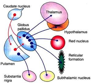
Connections of basal ganglia
FUNCTIONS OF BASAL GANGLIA The basal ganglia form the part of extrapyramidal system, which is concerned with motor activities. The various functions of basal ganglia are:
Fig.31. Afferent connections of corpus striatum.
Fig.32. Efferent and intrinsic connections of corpus striatum.
1. CONTROL OF VOLUNTARY MOTOR ACTIVITY The movements during voluntary motor activity are initiated by cerebral cortex. However, these movements are controlled by basal ganglia, which are in close association with cerebral cortex. During lesions of basal ganglia, this controlling mechanism is lost and so movements become inaccurate and awkward. Basal ganglia control the motor activities because of the nervous (neuronal) circuits between basal ganglia and other parts of the brain involved in motor activity. The neuronal circuits arise from three areas of the cerebral cortex. 1. Premotor area, 2. Primary motor area and 3. Supplementary motor area All these nerve fibers from cerebral cortex reach the caudate nucleus. From here, the fibers go to putamen. Some of fibers from cerebral cortex go directly to putamen also. Putamen sends fibers to globus pallidus. The fibers from here run towards the thalamus, subthalamic nucleus of Luys and substantia nigra. The subthalamic nucleus and substantia nigra are in turn, projected into thalamus. Now, the fibers from thalamus are projected back into the primary motor area and the other two motor areas, i.e. premotor area and supplementary motor area. Fibers between cerebral cortex and caudate nucleus are concerned with regulation of conscious movements known as the cognitive control of activity. The cortical fibers reaching putamen are directly concerned with control of subconscious execution of some movements during performance of trained motor activities, i.e. skilled activities. 2. CONTROL OF MUSCLE TONE Gamma motor neurons of spinal cord are responsible for maintaining the tone of muscles, which is important for posture. The tone of the muscle also depends on actions of muscle spindle. Gamma motor neurons, muscle spindle and muscle tone are all controlled by basal ganglia especially substantia nigra. In the lesion of basal ganglia, tone increases leading to rigidity. 3. CONTROL OF REFLEX MUSCULAR ACTIVITY Some of the reflex muscular activities, particularly visual and labyrinthine reflexes are important in the maintenance of posture. Coordination and integration of impulses for these activities depend upon basal ganglia. During lesion of basal ganglia, postural movements, especially visual and labyrinthine reflexes become abnormal. These abnormal movements are associated with rigidity. Rigidity is because of the loss of inhibitory influence from the cerebral cortex on spinal cord via basal gangliа 4. CONTROL OF AUTOMATIC ASSOCIATED MOVEMENTS Automatic associated movements are the movements in body, which take place along with some motor activities. Examples are the swing of the arms while walking, appropriate facial expressions while talking or doing any work. Basal ganglia are responsible for these movements. The lesion in basal ganglia causes absence of these automatic associated movements, resulting in poverty of movements. Face without appropriate expressions while doing any work is called mask like face. Body without associated movements is called statue like body.
5. ROLE IN AROUSAL (EXCITIVE) MECHANISM Globus pallidus and red nucleus are involved in arousal mechanism because of their connections with reticular formation. Extensive lesion in globus pallidus causes drowsiness, leading to sleep.
ROLE OF NEUROTRANSMITTERS IN THE FUNCTIONS OF BASAL GANGLIA The functions of basal ganglia on motor activities are executed by some neurotransmitters released by nerve endings within basal ganglia. Following neurotransmitters are released in basal ganglia. Dopamine: It is released by dopaminergic fibers from substantia nigra to corpus striatum (putamen and caudate nucleus - nigra strial fibers). The deficiency of dopamine leads to Parkinsonism. Gamma aminobutyric acid (GABA): It is secreted by intrinsic fibers of corpus striatum and substantia nigra. Acetylcholine: It is released by fibers from cerebral cortex to caudate nucleus and putamen. Substance P and enkephalins: These are released by fibers from globus pallidus reaching substantia nigra. Noradrenaline: This is secreted by the fibers between basal ganglia and reticular formation. Among all these neurotransmitters, dopamine and GABA are inhibitory neurotransmitters. So, the dopaminergic fibers and the fibers releasing GABA are inhibitory fibers. All other transmitters possess excitatory function.
|
|||||||||||||||||
|
|
Последнее изменение этой страницы: 2021-03-09; просмотров: 238; Нарушение авторского права страницы; Мы поможем в написании вашей работы! infopedia.su Все материалы представленные на сайте исключительно с целью ознакомления читателями и не преследуют коммерческих целей или нарушение авторских прав. Обратная связь - 3.144.103.10 (0.005 с.) |