Заглавная страница Избранные статьи Случайная статья Познавательные статьи Новые добавления Обратная связь КАТЕГОРИИ: ТОП 10 на сайте Приготовление дезинфицирующих растворов различной концентрацииТехника нижней прямой подачи мяча. Франко-прусская война (причины и последствия) Организация работы процедурного кабинета Смысловое и механическое запоминание, их место и роль в усвоении знаний Коммуникативные барьеры и пути их преодоления Обработка изделий медицинского назначения многократного применения Образцы текста публицистического стиля Четыре типа изменения баланса Задачи с ответами для Всероссийской олимпиады по праву 
Мы поможем в написании ваших работ! ЗНАЕТЕ ЛИ ВЫ?
Влияние общества на человека
Приготовление дезинфицирующих растворов различной концентрации Практические работы по географии для 6 класса Организация работы процедурного кабинета Изменения в неживой природе осенью Уборка процедурного кабинета Сольфеджио. Все правила по сольфеджио Балочные системы. Определение реакций опор и моментов защемления |
Task 1. Galwani’s first experiment.
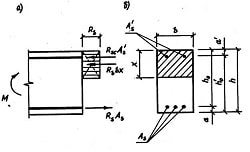
The investigator must destroy frog’s spine with preparation needle, destroying body in 2 cm in front of articulation place of spine and pelvis bones. It’s necessary to remove frontal body part and abdomen wall with visceral organs, to remove skin from posterior legs. Copper hook of “balcony” is bought to lumbo-sacral plexus radixes (rootlets, radicles). For this aim the investigator must hang the preparation on it. Then one should touch the legs muscles with “balcony” zinc plate. In the touching moment all legs are contracted. Draw the experiment scheme in your copy-book. Explain the reason of bimetallic “balcony” irritative action. Task 2. Galwani’s second experiment (contraction without metal). To prepare the preparation “sciatic nerve-legs muscles”. To catch the spine residue with plastic tweezers, not touching the nerve. On femur muscles resting after the preparation one must make transversal cutting and throw on the nerve on it for the nerve’s touching to the injured electronegative and non-injured electropositive muscle locuses. The experiment must be carried out some times while observing under the preparation muscles. It’s very important to take the nerve possessing high excitability. Draw the experiment scheme in your copy-books using the next figure. Second Galwani’s experiment scheme. Explain the reason of preparation muscles contraction. Task 3. K.Matteuchi’s experiment. To prepare 2 nervo-muscular preparations “sciatic nerve-legs muscles”. Put them to the dry dielectric plate so that the first preparation nerve were touching to the current source electrodes and the second preparation nerve must be lied longitudinally to the first preparation muscles. After that one should act to the first preparation sciatic nerve with unconstant current in course of some seconds. The result of the current action: both legs musclular contractions. The second legs muscular contractions (the nerve of which are located on the first preparation muscle) are called secondary. Draw the experiment’s scheme in your copy-book. They say traditionally that the second preparation nerve irritation reason is the first preparation skeletal muscle action currents.
5. Literature recommended: 1. Lecture course. 2. Mistchenko V.P., Tkachenko E.V. Methodical instructions for dental students (short lecture course).-Poltava, 2005.-P. 3-4. 3. Mistchenko V.P., Tkachenko E.V. Methodical instructions for medical students (short lecture course).-Poltava, 2005.-P. 4-5. 4. Ganong W.F. Review of Medical Physiology.-21st ed.-2003.-Section II. 5. Kapit W., Macey R.I., Meisami E. The Physiology Colouring Book: Harpers Collins Publishers, 1987.-P. 8-11, 14. 6. Bullock J., Boyle III J., Wang M.B. Physiology.-1991.-. P. 7. Guyton – Ganong – Chatterjee. Concise Physiology /Ed. By Dr Raja Shahzad Gull: M.B.B.S., F.C.P.S., King Edward Medical College.-Lahore, 1998 (1st Edition).-P.10-19. 8. Guyton A.C. Textbook of Medical Physiology.-NY, 1992.-P.88-113.
6. Materials for self-control: A. Control questions. 1. Bioelectrical phenomena investigation methods. 2. Resting potential: a) Appearence reasons and ion gradients levels. Potassium-sodium pump. b) Plasmatic membrane permeability for different ions. c) Membrane potential appearence mechanisms. Membrane potential level. 3. Action potential: a) Action potential appearence conditions and reasons. Local answer. Depolari- zation critical level. b) Action potential altitude and duration. Rule “everything or nothing”. c) Action potential appearence and development mechanism. Action potential phases. 4. Bioelectrical phenomena registration practical significance.
LESSON 3 1. The topic studied actuality. This functional method has the most spread usage in neurology and dentistry. EMG can show not only peripheral neuron damage but also big brain (brain cortex) structures pathological changings.
EMG can be performed at different muscles states: – at their relaxation; – at reflectory tonus changes (during other muscles tension, under emotional reactions, at deep inspiration); – during arbitrary contractions. Electromyogram is useful in the diagnosis of such neuro-muscular diseases as: 1.Motor neuron lesions 2.Peripheral nerve injury 3.Myotonia and 4.Myasthenia gravis Generally, myopathy means disease of skeletal muscles. Myopathies may be acquired or genetically derived. These diseases may or may not involve the nervous system. The common diseases of skeletal muscles are: 1.Muscular dystrophy 2.Diseases involving muscle tone 3.Fibrillation and denervation hypersensitivity and 4.Myasthenia gravis. І. MUSCULAR DYSTROPHY Muscular dystrophy is a disease characterized by progressive degeneration of muscle fibers without the involvement of nervous system. Mostly it has a hereditary origin. Common types of muscular dystrophy are Duchenne muscular dystrophy and Becker's muscular dystrophy. Duchenne Muscular Dystrophy This is an inherited sex-linked recessive disorder due to the absence of a gene product called dystrophin in the X chromosome. Dystrophin is necessary for the stability of sarcolemma. This disease is characterized by degeneration and necrosis of muscle fibers. The degenerated muscle fibers are replaced by fat and fibrous tissue. The common symptom is the muscular weakness. Sometimes there is enlargement of muscles (pseudohypertrophy). In severe conditions, respiratory muscles become weak resulting in difficulty in breathing. Becker's Muscular Dystrophy This is also inherited as sex-linked disorder. It is due to the reduction in quantity or alteration of dystrophin. The common features of this disorder are slow progressive weakness of legs and pelvis, pseudohypertrophy of calf muscles, difficulty in walking, fatigue and mental retardation. II. DISEASES INVOLVING MUSCLE TONE Hypotonicity This is the condition with decreased tone in the voluntary muscles. It is due to sectioning of spinal motor nerve to a muscle (lower motor neuron lesion). The tone of the muscle is decreased or lost. The muscle offers very little resistance to stretch. This is called flaccidity. The paralysis of muscle with hypotonicity is called flaccid paralysis. Hypertonicity This is characterized by increased muscle tone. It occurs in upper motor neuron lesion. During the lesion of upper motor neuron, the gamma motor neurons in the spinal cord are not inhibited. So, the discharge from these neurons is exaggerated and the tone of the muscle is increased. The muscle offers high resistance to stretch. This property is called spasticity. Myotonia This is an inherited disease characterized by continuous contraction of muscle even after the cessation of voluntary act. The capacity to relax is decreased. Myotonia is due to some abnormal gene that affects the ionic channels in sarcolemma.
|
|||||
|
|
Последнее изменение этой страницы: 2021-03-09; просмотров: 50; Нарушение авторского права страницы; Мы поможем в написании вашей работы! infopedia.su Все материалы представленные на сайте исключительно с целью ознакомления читателями и не преследуют коммерческих целей или нарушение авторских прав. Обратная связь - 3.139.83.178 (0.007 с.) |