Заглавная страница Избранные статьи Случайная статья Познавательные статьи Новые добавления Обратная связь КАТЕГОРИИ: ТОП 10 на сайте Приготовление дезинфицирующих растворов различной концентрацииТехника нижней прямой подачи мяча. Франко-прусская война (причины и последствия) Организация работы процедурного кабинета Смысловое и механическое запоминание, их место и роль в усвоении знаний Коммуникативные барьеры и пути их преодоления Обработка изделий медицинского назначения многократного применения Образцы текста публицистического стиля Четыре типа изменения баланса Задачи с ответами для Всероссийской олимпиады по праву 
Мы поможем в написании ваших работ! ЗНАЕТЕ ЛИ ВЫ?
Влияние общества на человека
Приготовление дезинфицирующих растворов различной концентрации Практические работы по географии для 6 класса Организация работы процедурного кабинета Изменения в неживой природе осенью Уборка процедурного кабинета Сольфеджио. Все правила по сольфеджио Балочные системы. Определение реакций опор и моментов защемления |
Image S21: Pelger Huet neutrophils ⇐ ПредыдущаяСтр 3 из 3
Pelger Huet anomaly – classic bi-lobed cells with dense chromatin condensation but normal granulation
J. Burthem, M. Brereton
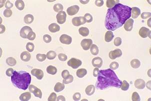
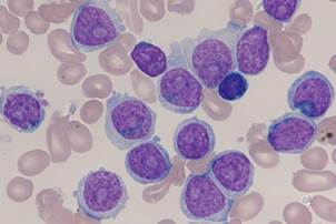
Image S22: leukaemic myeloblasts
Acute myeloid leukaemia (AML) – hypogranular primitive blast cells
J. Burthem, M. Brereton
Image S23: abnormal promyelocytes in APL (1)
APML – two hypergranular promyelocytes
J. Burthem, M. Brereton
Image S24: abnormal promyelocytes in APL (2)
Abnormal promyelocyte containing multiple Auer rods (faggot cell)
G. Rozenberg
(Copyright: Microscopic haematology: a practical guide for the laboratory 3e (c) 2011, Sydney, Elsevier Australia) 7
Supplementary images: ICSH Recommendations for Peripheral Blood Cell Morphology
Standardization and Grading
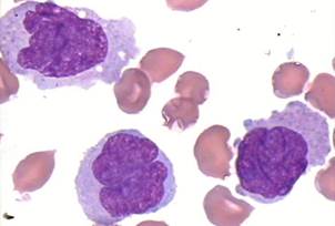
Image S25: monoblasts
Acute monoblastic leukaemia – monoblasts and promonocytes
J. Burthem, M. Brereton
Image S26: abnormal promonocytes
Chronic myelomonocytic leukaemia (CMML)
G. Zini
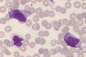
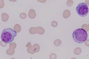
Image S27: reactive lymphocytes
Infectious mononucleosis – typical reactive lymphocytes with flowing basophilic cytoplasm
J. Burthem, M. Brereton
Image S28: hairy cells
Hairy cell leukaemia
J. Burthem, M. Brereton 8
Supplementary images: ICSH Recommendations for Peripheral Blood Cell Morphology
Standardization and Grading
Image S29: follicular lymphoma cells
Circulating follicular lymphoma cells – note the small cells with cleaved nuclei and sparse cytoplasm
J. Burthem, M. Brereton
Image S30: plasma cells
Plasma cell leukaemia. Note also the background protein staining and the associated red cell rouleaux. Note that one plasma cell has features of immaturity and may be regarded as a plasmablast.
J. Burthem, M. Brereton
Image S31: prolymphocytic leukaemia cells
B-Prolymphocytic leukaemia
J. Burthem, M. Brereton
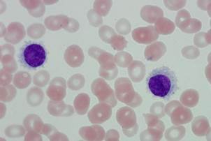
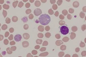
Image S32: chronic lymphocytic leukaemia cells
Typical CLL lymphocytes with a smudge cell
J. Burthem, M. Brereton 9
Supplementary images: ICSH Recommendations for Peripheral Blood Cell Morphology
Standardization and Grading
Image S33: giant platelets
Myelofibrosis – some large and giant abnormally granulated platelets and a micromegakaryocyte
J. Burthem, M. Brereton
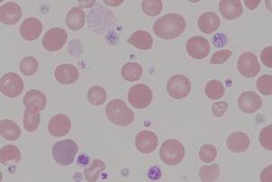
Image S34: hypogranular platelets
Myelofibrosis – variable platelet appearance with normal and abnormally granulated platelets together with some large hypogranular forms
J. Burthem, M. Brereton
Image S35: micromegakaryocytes
Myelofibrosis – note the typical ‘granulated’ platelet cytoplasm. A bare megakaryocyte nucleus is also present
J. Burthem, M. Brereton
|
||||
|
|
Последнее изменение этой страницы: 2021-03-09; просмотров: 219; Нарушение авторского права страницы; Мы поможем в написании вашей работы! infopedia.su Все материалы представленные на сайте исключительно с целью ознакомления читателями и не преследуют коммерческих целей или нарушение авторских прав. Обратная связь - 3.138.179.119 (0.006 с.) |